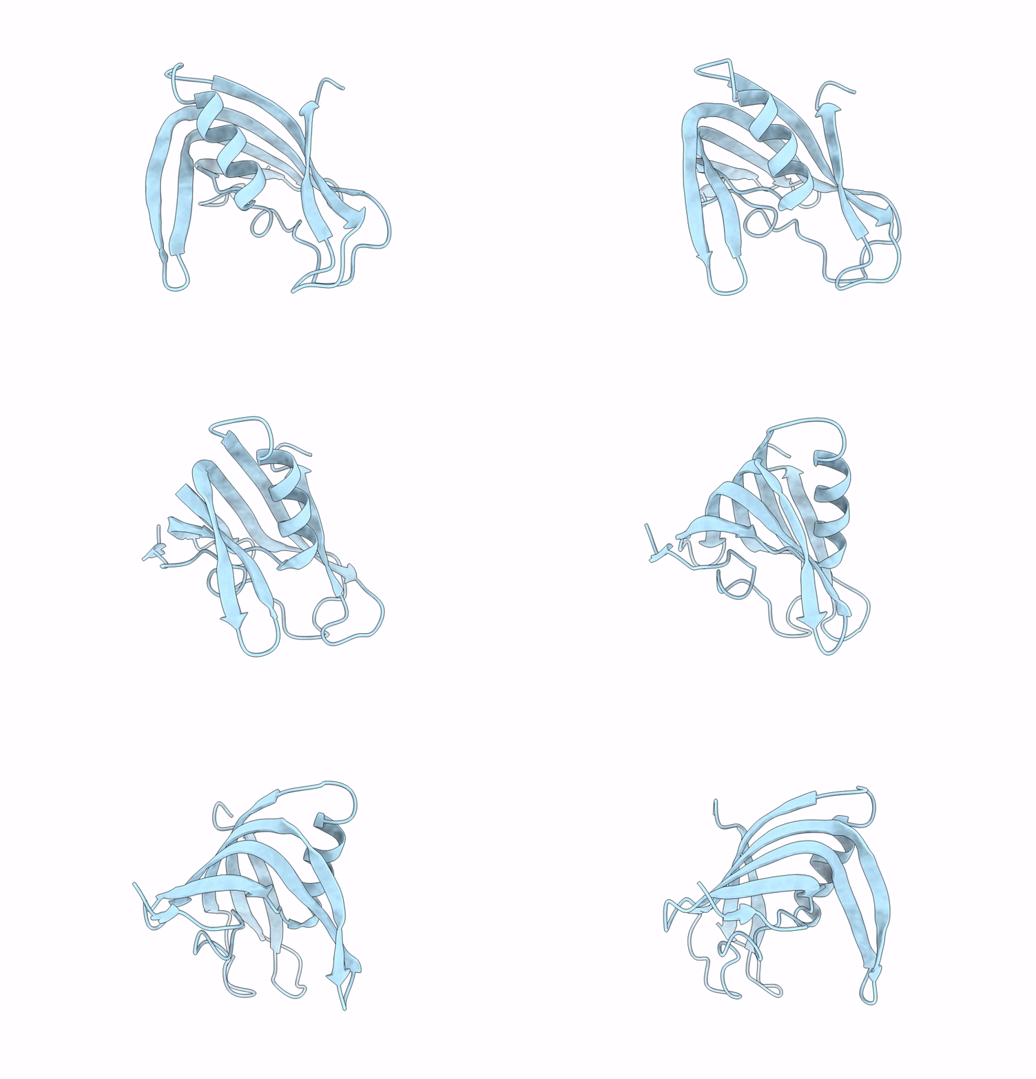
9GFQ image
Entry Detail
PDB ID:
9GFQ
Title:
Structure of PRD1 SSB P12
Biological Source:
Source Organism:
Host Organism:
PDB Version:
Deposition Date:
2024-08-12
Release Date:
2025-04-09
Method Details:
Experimental Method:
Resolution:
1.70 Å
R-Value Free:
0.25
R-Value Work:
0.22
Space Group:
P 21 21 21
Macromolecular Entities
Polymer Type:polypeptide(L)
Description:Single-stranded DNA-binding protein
Chain IDs:A
Chain Length:160
Number of Molecules:1
Biological Source:Enterobacteria phage PRD1
Primary Citation
Structural basis for cooperative ssDNA binding by bacteriophage protein filament P12.
Nucleic Acids Res. 53 ? ? (2025)
PMID: 40052821 DOI: 10.1093/nar/gkaf132

Abstact

Protein-primed DNA replication is a unique mechanism, bioorthogonal to other known DNA replication modes. It relies on specialised single-stranded DNA (ssDNA)-binding proteins (SSBs) to stabilise ssDNA intermediates by unknown mechanisms. Here, we present the structural and biochemical characterisation of P12, an SSB from bacteriophage PRD1. High-resolution cryo-electron microscopy reveals that P12 forms a unique, cooperative filament along ssDNA. Each protomer binds the phosphate backbone of 6 nucleotides in a sequence-independent manner, protecting ssDNA from nuclease degradation. Filament formation is driven by an intrinsically disordered C-terminal tail, facilitating cooperative binding. We identify residues essential for ssDNA interaction and link the ssDNA-binding ability of P12 to toxicity in host cells. Bioinformatic analyses place the P12 fold as a distinct branch within the OB-like fold family. This work offers new insights into protein-primed DNA replication and lays a foundation for biotechnological applications.

Legend

Protein

Chemical

Disease

Primary Citation of related structures