Deposition Date
2024-07-26
Release Date
2025-06-04
Last Version Date
2025-06-04
Entry Detail
PDB ID:
9GA6
Keywords:
Title:
The crystal structure of human Annexin A4 derived from crystals grown in 40 mM of CaCl2
Biological Source:
Source Organism(s):
Homo sapiens (Taxon ID: 9606)
Expression System(s):
Method Details:
Experimental Method:
Resolution:
1.27 Å
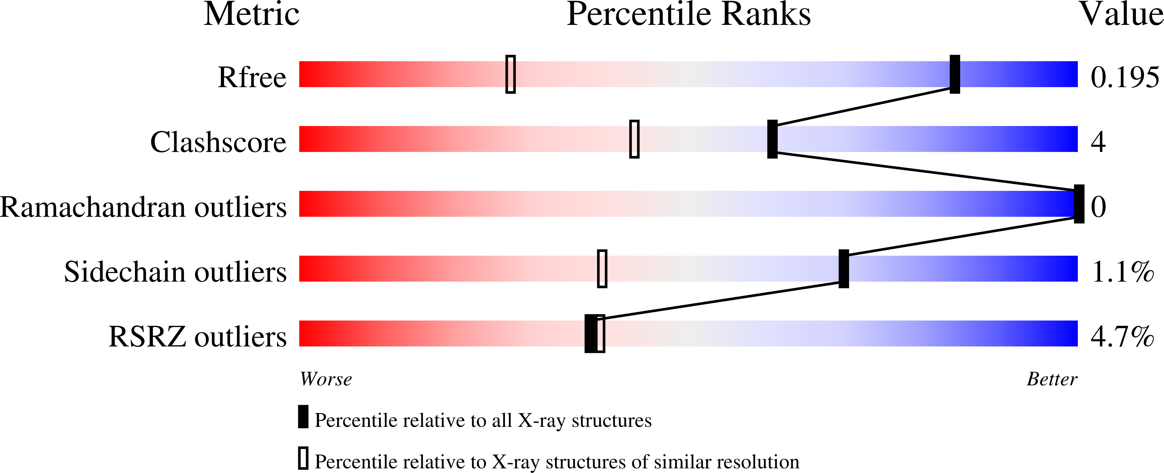
R-Value Free:
0.18
R-Value Work:
0.17
Space Group:
P 1 21 1