Deposition Date
2024-07-22
Release Date
2025-05-21
Last Version Date
2025-11-26
Entry Detail
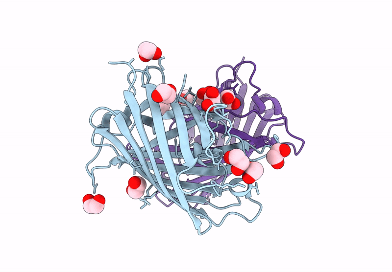
Biological Source:
Source Organism(s):
Cytaeis uchidae (Taxon ID: 1254443)
Expression System(s):
Method Details:
Experimental Method:
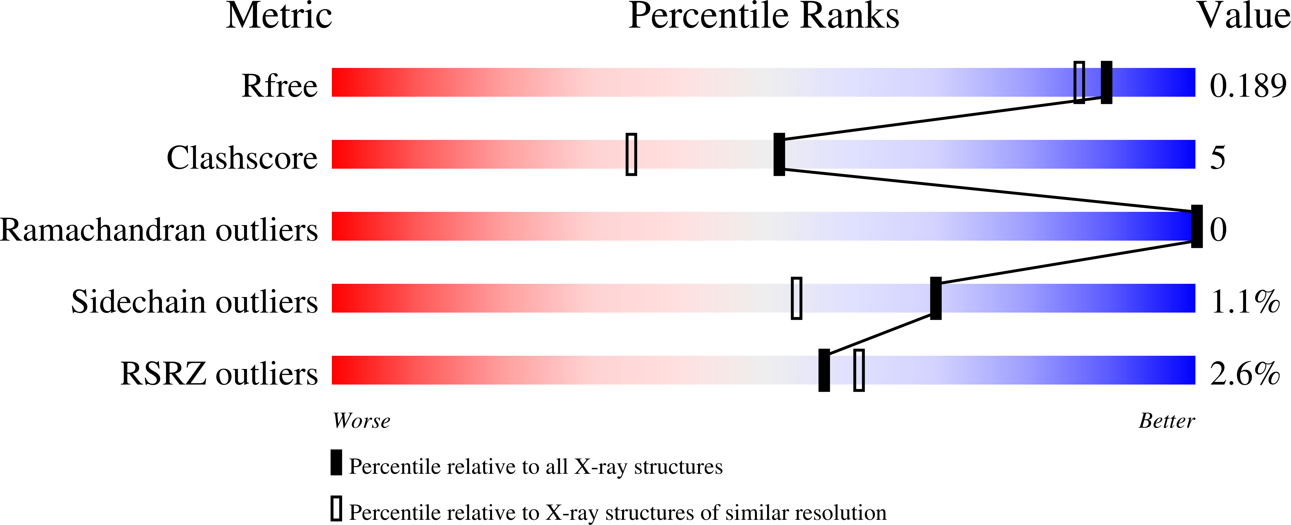
Resolution:
1.65 Å
R-Value Free:
0.17
R-Value Work:
0.15
Space Group:
P 61