9G0B image
Deposition Date 2024-07-07
Release Date 2024-07-31
Last Version Date 2025-10-29
Entry Detail
PDB ID:
9G0B
Keywords:
Title:
Rhinovirus A2 uncoating intermediate revealing the natural pocket factor (pH 5.8 and 4 degrees Celsius)
Biological Source:
Source Organism:
rhinovirus A2 (Taxon ID: 12130)
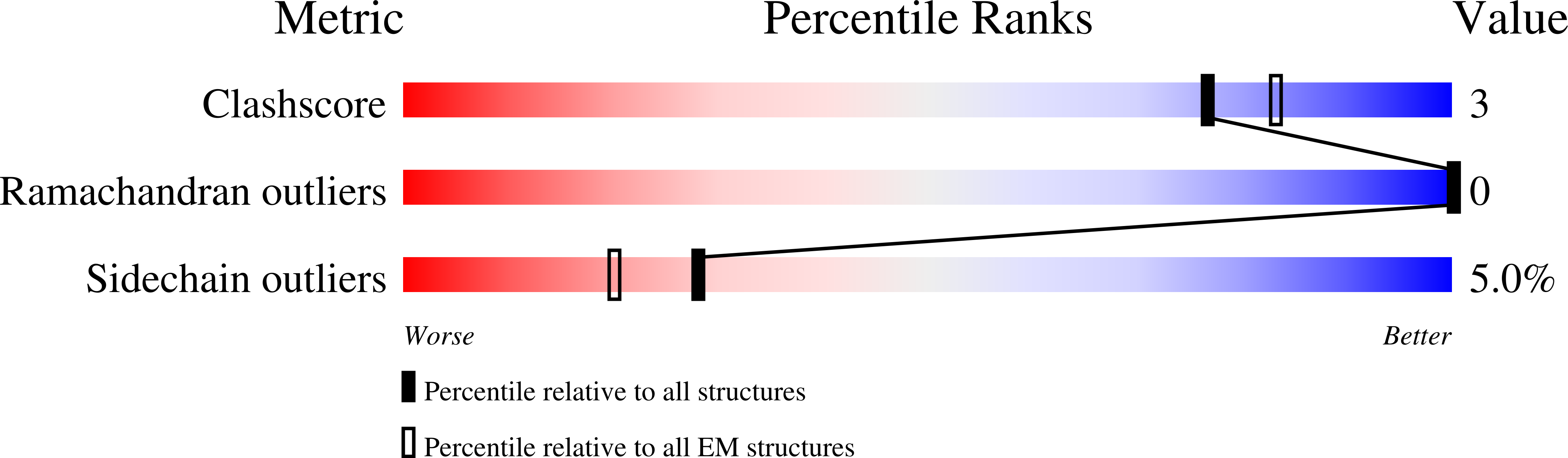
Method Details:
Experimental Method:
Resolution:
3.20 Å
Aggregation State:
PARTICLE
Reconstruction Method:
SINGLE PARTICLE
Macromolecular Entities
Structures with similar UniProt ID
Protein Blast
Polymer Type:polypeptide(L)
Molecule:Capsid protein VP1
Chain IDs:A
Chain Length:283
Number of Molecules:1
Biological Source:rhinovirus A2
Structures with similar UniProt ID
Protein Blast
Polymer Type:polypeptide(L)
Molecule:Capsid protein VP2
Chain IDs:B
Chain Length:261
Number of Molecules:1
Biological Source:rhinovirus A2
Structures with similar UniProt ID
Protein Blast
Polymer Type:polypeptide(L)
Molecule:Capsid protein VP3
Chain IDs:C
Chain Length:237
Number of Molecules:1
Biological Source:rhinovirus A2
Structures with similar UniProt ID
Protein Blast
Polymer Type:polypeptide(L)
Molecule:Capsid protein VP4
Chain IDs:D
Chain Length:68
Number of Molecules:1
Biological Source:rhinovirus A2
Ligand Molecules
Primary Citation
New rhinovirus uncoating intermediate reveals how sodium versus potassium ions influence RNA release.
Sci Rep 15 36768 36768 (2025)
PMID: 41120457 DOI: 10.1038/s41598-025-20627-0

Abstact

Electron microscopy (EM) of rhinovirus A2 (RV-A2) incubated in Na+ phosphate buffer (pH 7.6) for 12 h at 25 °C revealed partial fragmentation, whereas upon incubation in K+ phosphate buffer, RV-A2 appeared intact. In buffers adjusted to pH 5.8, these differences became more pronounced; acidic Na+ phosphate buffer promoted disintegration of the particles, whereas in acidic K+ phosphate buffer, the virus appeared like native. Incubation in the acidic buffers for one hour at 4 °C followed by neutralisation resulted in the respective formation of non-infectious A particles (in Na+) and a non-infectious novel uncoating intermediate (in K+), which we termed 'E0 particle'. Negative staining EM revealed phosphotungstate penetration into A particles, but not into E0 particles. Cryo-EM image reconstruction of the E0 particle showed clear differences between A and E0 particles; like native virus, E0 contained VP4 and a pocket factor. Native RV-A2 RNA cores, obtained by gentle proteinase-K digestion in K+ and Na+ phosphate buffer, respectively, differed in accessibility of dsRNA regions, detected by PaSTRy. Variance in RNA compactness observed in K+ versus Na+ phosphate buffer was confirmed by rotary shadowing EM; in K+ phosphate buffer, the RNA remained condensed while, in Na+ phosphate buffer, distinct unfolding stages were apparent.

Legend

Protein

Chemical

Disease

Primary Citation of related structures