9FVR image
Entry Detail
PDB ID:
9FVR
Title:
Transcription repressor NrdR from E. coli, ATP/dATP-bound state, SeMet protein
Biological Source:
Source Organism:
Host Organism:
PDB Version:
Deposition Date:
2024-06-28
Release Date:
2025-03-19
Method Details:
Experimental Method:
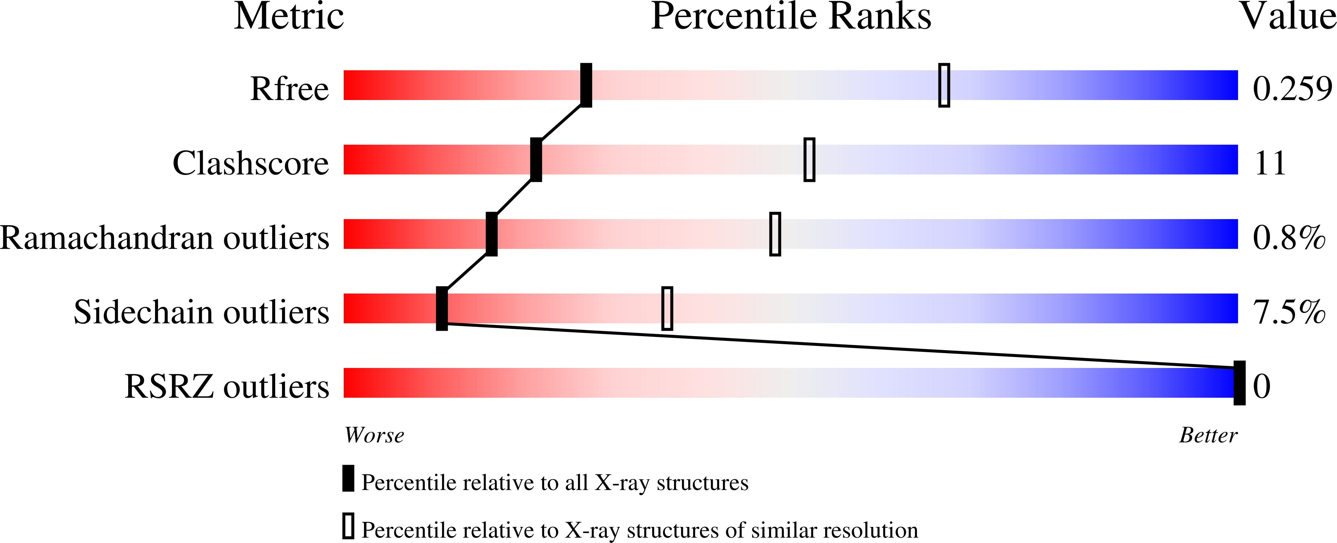
Resolution:
3.10 Å
R-Value Free:
0.27
R-Value Work:
0.22
R-Value Observed:
0.22
Space Group:
P 21 21 21
Macromolecular Entities
Polymer Type:polypeptide(L)
Description:Transcriptional repressor NrdR
Chain IDs:A, B, C, D
Chain Length:155
Number of Molecules:4
Biological Source:Escherichia coli
Modified Residue
Compound ID Chain ID Parent Comp ID Details 2D Image
MSE A MET modified residue
Primary Citation
Bacterial transcriptional repressor NrdR - a flexible multifactorial nucleotide sensor.
Febs J. ? ? ? (2025)
PMID: 40029022 DOI: 10.1111/febs.70037

Abstact

NrdR is a bacterial transcriptional repressor consisting of a zinc (Zn)-ribbon domain followed by an ATP-cone domain. Understanding its mechanism of action could aid the design of novel antibacterials. NrdR binds specifically to two "NrdR boxes" upstream of ribonucleotide reductase operons, of which Escherichia coli has three: nrdHIEF, nrdDG and nrdAB, in the last of which we identified a new box. We show that E. coli NrdR (EcoNrdR) has similar binding strength to all three sites when loaded with ATP plus deoxyadenosine triphosphate (dATP) or equivalent diphosphate combinations. No other combination of adenine nucleotides promotes binding to DNA. We present crystal structures of EcoNrdR-ATP-dATP and EcoNrdR-ADP-dATP, which are the first high-resolution crystal structures of an NrdR. We have also determined cryo-electron microscopy structures of DNA-bound EcoNrdR-ATP-dATP and novel filaments of EcoNrdR-ATP. Tetrameric forms of EcoNrdR involve alternating interactions between pairs of Zn-ribbon domains and ATP-cones. The structures reveal considerable flexibility in relative orientation of ATP-cones vs Zn-ribbon domains. The structure of DNA-bound EcoNrdR-ATP-dATP shows that significant conformational rearrangements between ATP-cones and Zn-ribbons accompany DNA binding while the ATP-cones retain the same relative orientation. In contrast, ATP-loaded EcoNrdR filaments show rearrangements of the ATP-cone pairs and sequester the DNA-binding residues of NrdR such that they are unable to bind to DNA. Our results, in combination with a previous structural and biochemical study, point to highly flexible EcoNrdR structures that, when loaded with the correct nucleotides, adapt to an optimal promoter-binding conformation.

Legend

Protein

Chemical

Disease

Primary Citation of related structures