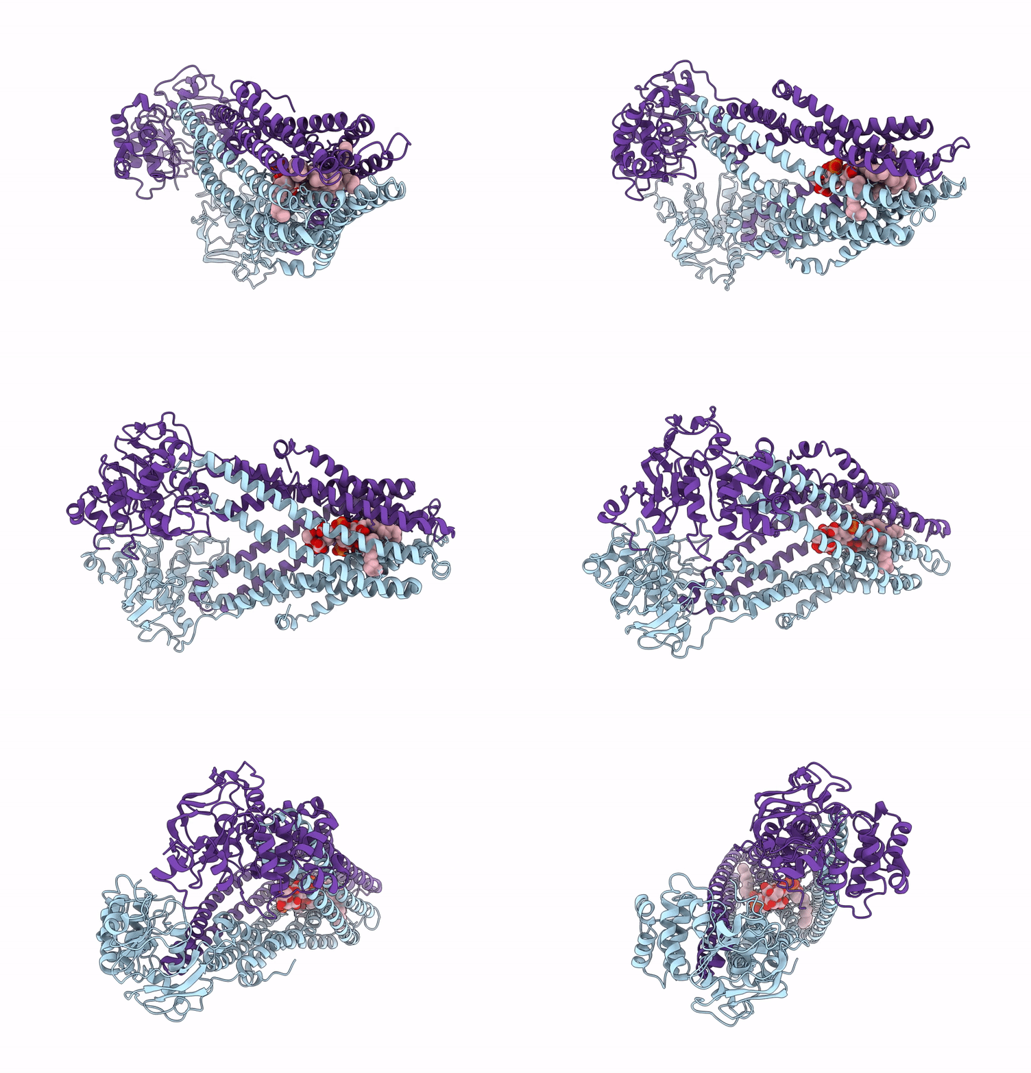
9FV5 image
Deposition Date 2024-06-26
Release Date 2025-03-19
Last Version Date 2025-07-09
Entry Detail
PDB ID:
9FV5
Title:
MsbA in peptidisc inward-facing narrow open
Biological Source:
Source Organism:
Escherichia coli (Taxon ID: 562)
Host Organism:
Method Details:
Experimental Method:
Resolution:
3.90 Å
Aggregation State:
PARTICLE
Reconstruction Method:
SINGLE PARTICLE
Macromolecular Entities
Polymer Type:polypeptide(L)
Molecule:ATP-dependent lipid A-core flippase
Gene (Uniprot):msbA
Chain IDs:A, B
Chain Length:582
Number of Molecules:2
Biological Source:Escherichia coli
Ligand Molecules
Primary Citation
The ABC transporter MsbA in a dozen environments.
Structure 33 916 ? (2025)
PMID: 40056915 DOI: 10.1016/j.str.2025.02.002

Abstact

High-resolution structure determination of membrane proteins typically requires reconstitution into artificial membrane mimics. The choice of the specific membrane substitute can strongly affect the protein's specific activity, stability, and conformational spectrum, potentially leading to errors or misinterpretation during analysis. The bacterial ATP-binding cassette transporter MsbA is a prominent example of such environment-specific bias. Here, we present a systematic analysis of the conformational spectrum of MsbA, stabilized in a dozen environments, using cryoelectron microscopy (cryo-EM), and show pronounced feedback between the membrane mimetics and the transporter. Detergents generally favor wide inward-facing conformations while nanodiscs induce narrower conformations. Notably, only in three tested environments, MsbA samples the full movement of the nucleotide-binding domains, including narrow and wide conformations. We expect this study to serve as a blueprint for other membrane proteins, even where a structural reaction to the hydrophobic environment is not directly visible but still critical for the proteins' function.

Legend

Protein

Chemical

Disease

Primary Citation of related structures