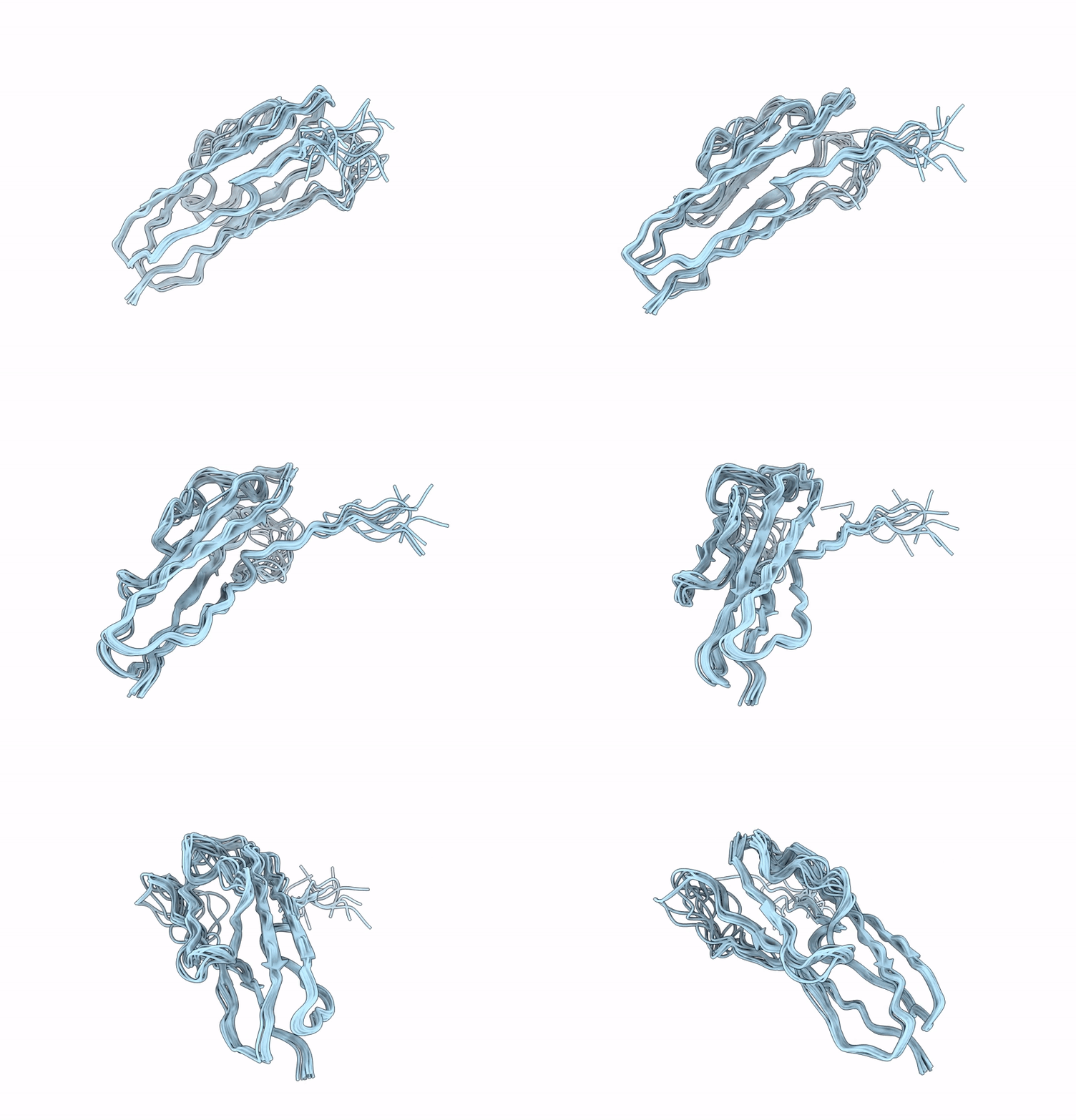
9FU8 image
Deposition Date 2024-06-26
Release Date 2025-04-02
Last Version Date 2025-04-02
Entry Detail
PDB ID:
9FU8
Title:
Structure of the ASH1 domain of Drosophila melanogaster Spd-2
Biological Source:
Source Organism:
Host Organism:
Method Details:
Experimental Method:
Conformers Calculated:
100
Conformers Submitted:
10
Selection Criteria:
target function
Macromolecular Entities
Polymer Type:polypeptide(L)
Molecule:BcDNA.LD24702
Gene (Uniprot):spd-2
Chain IDs:A
Chain Length:113
Number of Molecules:1
Biological Source:Drosophila melanogaster
Ligand Molecules
Primary Citation
The conserved Spd-2/CEP192 domain adopts a unique protein fold to promote centrosome scaffold assembly.
Sci Adv 11 eadr5744 eadr5744 (2025)
PMID: 40106572 DOI: 10.1126/sciadv.adr5744

Abstact

Centrosomes form when centrioles assemble pericentriolar material (PCM) around themselves. Spd-2/CEP192 proteins, defined by a conserved "Spd-2 domain" (SP2D) comprising two closely spaced AspM-Spd-2-Hydin (ASH) domains, play a critical role in centrosome assembly. Here, we show that the SP2D does not target Drosophila Spd-2 to centrosomes but rather promotes PCM scaffold assembly. Crystal structures of the human and honeybee SP2D reveal an unusual "extended cradle" structure mediated by a conserved interaction interface between the two ASH domains. Mutations predicted to perturb this interface, including a human mutation associated with male infertility and Mosaic Variegated Aneuploidy, disrupt PCM scaffold assembly in flies. The SP2D is monomeric in solution, but the Drosophila SP2D can form higher-order oligomers upon phosphorylation by PLK1 (Polo-like kinase 1). Crystal-packing interactions and AlphaFold predictions suggest how SP2Ds might self-assemble, and mutations associated with one such potential dimerization interface markedly perturb SP2D oligomerization in vitro and PCM scaffold assembly in vivo.

Legend

Protein

Chemical

Disease

Primary Citation of related structures