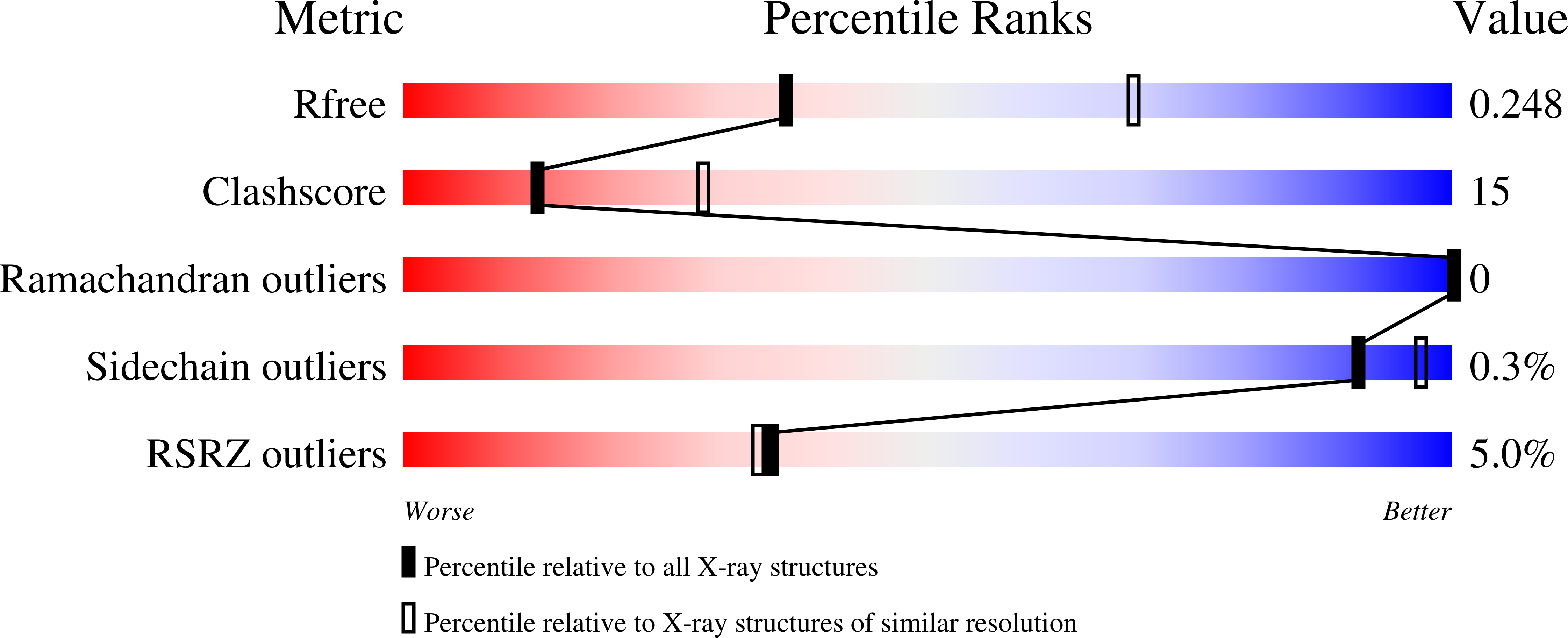
9FOI image
Deposition Date 2024-06-11
Release Date 2025-06-25
Last Version Date 2025-06-25
Entry Detail
PDB ID:
9FOI
Keywords:
Title:
Structure of human KCTD1
Biological Source:
Source Organism:
Homo sapiens (Taxon ID: 9606)
Host Organism:
Method Details:
Experimental Method:
Resolution:
2.71 Å
R-Value Free:
0.24
R-Value Work:
0.20
R-Value Observed:
0.20
Space Group:
P 1 21 1
Macromolecular Entities
Polymer Type:polypeptide(L)
Molecule:BTB/POZ domain-containing protein KCTD1
Gene (Uniprot):KCTD1
Chain IDs:A, B, C, D, E
Chain Length:258
Number of Molecules:5
Biological Source:Homo sapiens
Primary Citation
A BTB extension and ion-binding domain contribute to the pentameric structure and TFAP2A binding of KCTD1.
Structure 32 1586 1593.e4 (2024)
PMID: 39191250 DOI: 10.1016/j.str.2024.07.023

Abstact

KCTD family proteins typically assemble into cullin-RING E3 ligases. KCTD1 is an atypical member that functions instead as a transcriptional repressor. Mutations in KCTD1 cause developmental abnormalities and kidney fibrosis in scalp-ear-nipple syndrome. Here, we present unexpected mechanistic insights from the structure of human KCTD1. Disease-causing mutation P20S maps to an unrecognized extension of the BTB domain that contributes to both its pentameric structure and TFAP2A binding. The C-terminal domain (CTD) shares its fold and pentameric assembly with the GTP cyclohydrolase I feedback regulatory protein (GFRP) despite lacking discernible sequence similarity. Most surprisingly, the KCTD1 CTD establishes a central channel occupied by alternating sodium and iodide ions that restrict TFAP2A dissociation. The elucidation of the structure redefines the KCTD1 BTB domain fold and identifies an unexpected ion-binding site for future study of KCTD1's function in the ectoderm, neural crest, and kidney.

Legend

Protein

Chemical

Disease

Primary Citation of related structures