9FDA image
Entry Detail
PDB ID:
9FDA
EMDB ID:
Keywords:
Title:
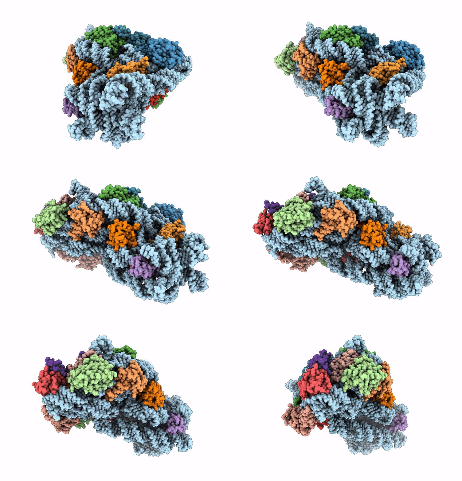
Structure of E. coli 30S-IF1-IF3-mRNA-Edeine complex
Biological Source:
Source Organism:
Host Organism:
PDB Version:
Deposition Date:
2024-05-16
Release Date:
2025-03-19
Method Details:
Experimental Method:
Resolution:
2.00 Å
Aggregation State:
PARTICLE
Reconstruction Method:
SINGLE PARTICLE
Macromolecular Entities
Polymer Type:polyribonucleotide
Description:16S rRNA
Chain IDs:O (auth: B)
Chain Length:1542
Number of Molecules:1
Biological Source:Escherichia coli
Polymer Type:polypeptide(L)
Description:Small ribosomal subunit protein uS4
Chain IDs:A (auth: D)
Chain Length:206
Number of Molecules:1
Biological Source:Escherichia coli
Polymer Type:polypeptide(L)
Description:Small ribosomal subunit protein uS5
Chain IDs:B (auth: E)
Chain Length:167
Number of Molecules:1
Biological Source:Escherichia coli
Polymer Type:polypeptide(L)
Description:Small ribosomal subunit protein bS6, fully modified isoform
Chain IDs:C (auth: F)
Chain Length:135
Number of Molecules:1
Biological Source:Escherichia coli
Polymer Type:polypeptide(L)
Description:Small ribosomal subunit protein uS8
Chain IDs:D (auth: H)
Chain Length:130
Number of Molecules:1
Biological Source:Escherichia coli
Polymer Type:polypeptide(L)
Description:Translation initiation factor IF-1
Chain IDs:E (auth: I)
Chain Length:72
Number of Molecules:1
Biological Source:Escherichia coli
Polymer Type:polypeptide(L)
Description:Translation initiation factor IF-3
Chain IDs:F (auth: J)
Chain Length:180
Number of Molecules:1
Biological Source:Escherichia coli
Polymer Type:polypeptide(L)
Description:Small ribosomal subunit protein uS11
Chain IDs:G (auth: K)
Chain Length:139
Number of Molecules:1
Biological Source:Escherichia coli
Polymer Type:polypeptide(L)
Description:Small ribosomal subunit protein uS12
Chain IDs:H (auth: L)
Chain Length:124
Number of Molecules:1
Biological Source:Escherichia coli
Polymer Type:polypeptide(L)
Description:Small ribosomal subunit protein uS15
Chain IDs:I (auth: O)
Chain Length:89
Number of Molecules:1
Biological Source:Escherichia coli
Polymer Type:polypeptide(L)
Description:Small ribosomal subunit protein bS16
Chain IDs:J (auth: P)
Chain Length:82
Number of Molecules:1
Biological Source:Escherichia coli
Polymer Type:polypeptide(L)
Description:Small ribosomal subunit protein uS17
Chain IDs:K (auth: Q)
Chain Length:84
Number of Molecules:1
Biological Source:Escherichia coli
Polymer Type:polypeptide(L)
Description:Small ribosomal subunit protein bS18
Chain IDs:L (auth: R)
Chain Length:75
Number of Molecules:1
Biological Source:Escherichia coli
Polymer Type:polypeptide(L)
Description:Small ribosomal subunit protein bS20
Chain IDs:M (auth: T)
Chain Length:87
Number of Molecules:1
Biological Source:Escherichia coli
Polymer Type:polypeptide(L)
Description:Small ribosomal subunit protein bS21
Chain IDs:N (auth: U)
Chain Length:71
Number of Molecules:1
Biological Source:Escherichia coli
Modified Residue
Compound ID Chain ID Parent Comp ID Details 2D Image
D2T H ASP modified residue
Primary Citation
The translation inhibitors kasugamycin, edeine and GE81112 target distinct steps during 30S initiation complex formation.
Nat Commun 16 2470 2470 (2025)
PMID: 40075065 DOI: 10.1038/s41467-025-57731-8

Abstact

During bacterial translation initiation, the 30S ribosomal subunit, initiation factors, and initiator tRNA define the reading frame of the mRNA. This process is inhibited by kasugamycin, edeine and GE81112, however, their mechanisms of action have not been fully elucidated. Here we present cryo-electron microscopy structures of 30S initiation intermediate complexes formed in the presence of kasugamycin, edeine and GE81112 at resolutions of 2.0-2.9 Å. The structures reveal that all three antibiotics bind within the E-site of the 30S and preclude 30S initiation complex formation. While kasugamycin and edeine affect early steps of 30S pre-initiation complex formation, GE81112 stalls pre-initiation complex formation at a further step by allowing start codon recognition, but impeding IF3 departure. Collectively, our work highlights how chemically distinct compounds binding at a conserved site on the 30S can interfere with translation initiation in a unique manner.

Legend

Protein

Chemical

Disease

Primary Citation of related structures