9FB1 image
Deposition Date 2024-05-11
Release Date 2024-12-18
Last Version Date 2024-12-18
Entry Detail
PDB ID:
9FB1
Keywords:
Title:
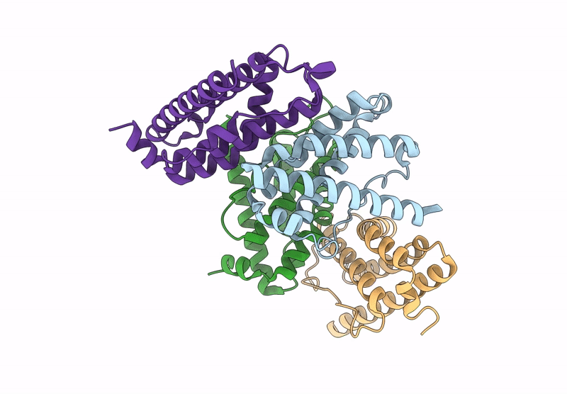
Crystal structure of Rv2242 regulator N-terminal fragment (1-160)
Biological Source:
Source Organism:
Method Details:
Experimental Method:
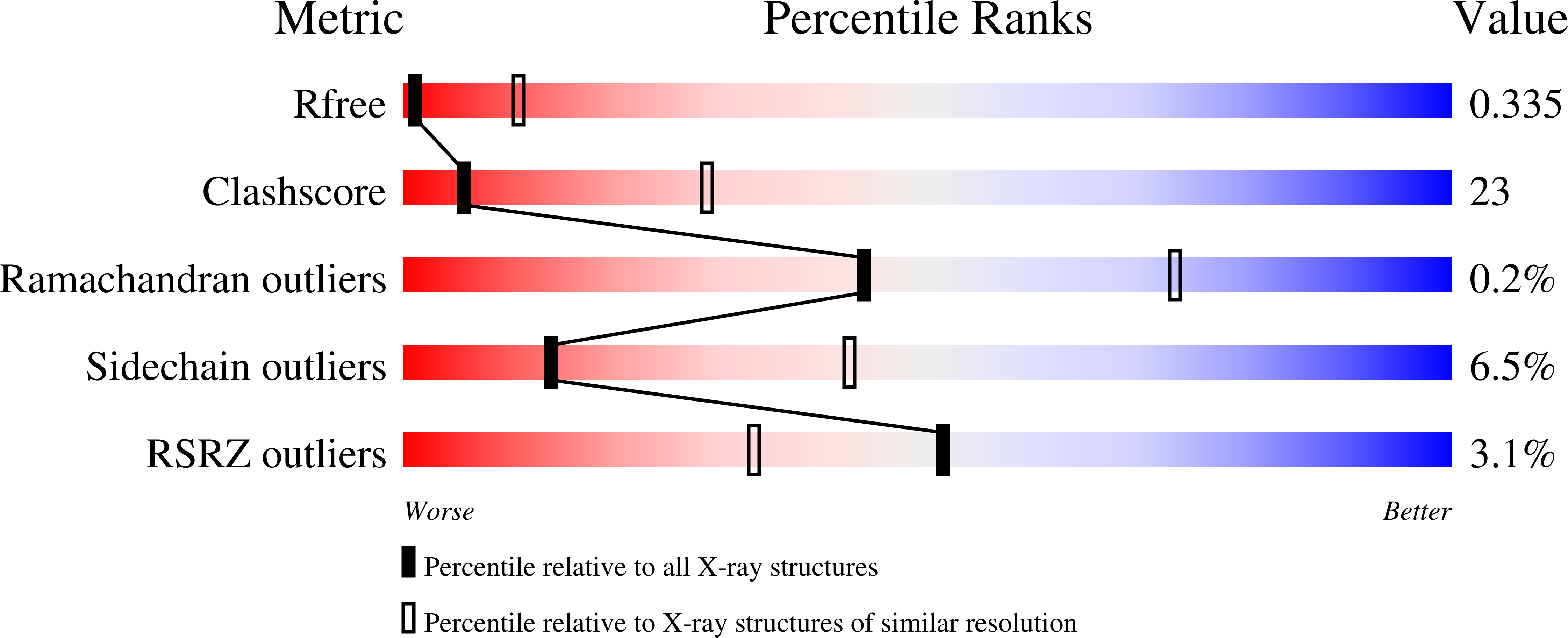
Resolution:
3.59 Å
R-Value Free:
0.32
R-Value Work:
0.25
R-Value Observed:
0.25
Space Group:
P 41 2 2
Macromolecular Entities
Polymer Type:polypeptide(L)
Molecule:Uncharacterized protein Rv2242
Gene (Uniprot):MTCY427.23
Chain IDs:A, B, C, D
Chain Length:181
Number of Molecules:4
Biological Source:Mycobacterium tuberculosis H37Rv
Ligand Molecules
Primary Citation
Domain architecture of the Mycobacterium tuberculosis MabR ( Rv2242 ), a member of the PucR transcription factor family.
Heliyon 10 e40494 e40494 (2024)
PMID: 39641026 DOI: 10.1016/j.heliyon.2024.e40494

Abstact

MabR (Rv2242), a PucR-type transcription factor, plays a crucial role in regulating mycolic acid biosynthesis in Mycobacterium tuberculosis. To understand its regulatory mechanisms, we determined the crystal structures of its N-terminal and C-terminal domains. The N-terminal domain adopts a globin-like fold, while the C-terminal domain comprises an α/β GGDEF domain and an all-α effector domain with a helix-turn-helix DNA-binding motif. This unique domain combination is specific to Actinomycetes. Biochemical and computational studies suggest that full-length MabR forms both dimeric and tetrameric assemblies in solution. Structural analysis revealed two distinct dimerization interfaces within the N- and C-terminal domains, further supporting a tetrameric organization. These findings provide valuable insights into the domain architecture, oligomeric state, and potential regulatory mechanisms of MabR.

Legend

Protein

Chemical

Disease

Primary Citation of related structures