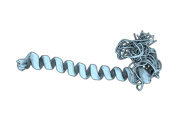
9F1G image
Deposition Date 2024-04-18
Release Date 2024-09-11
Last Version Date 2024-12-18
Entry Detail
PDB ID:
9F1G
Title:
Remorin protein StREM13_150-198
Biological Source:
Source Organism:
Method Details:
Experimental Method:
Conformers Calculated:
50
Conformers Submitted:
40
Selection Criteria:
structures with the least restraint violations
Macromolecular Entities
Polymer Type:polypeptide(L)
Molecule:Remorin
Chain IDs:A
Chain Length:49
Number of Molecules:1
Biological Source:Nicotiana benthamiana
Ligand Molecules
Primary Citation
Dynamic pre-structuration of lipid nanodomain-segregating remorin proteins.
Commun Biol 7 1620 1620 (2024)
PMID: 39639105 DOI: 10.1038/s42003-024-07330-y

Abstact

Remorins are multifunctional proteins, regulating immunity, development and symbiosis in plants. When associating to the membrane, remorins sequester specific lipids into functional membrane nanodomains. The multigenic protein family contains six groups, classified upon their protein-domain composition. Membrane targeting of remorins occurs independently from the secretory pathway. Instead, they are directed into different nanodomains depending on their phylogenetic group. All family members contain a C-terminal membrane anchor and a homo-oligomerization domain, flanked by an intrinsically disordered region of variable length at the N-terminal end. We here combined molecular imaging, NMR spectroscopy, protein structure calculations and advanced molecular dynamics simulation to unveil a stable pre-structuration of coiled-coil dimers as nanodomain-targeting units, containing a tunable fuzzy coat and a bar code-like positive surface charge before membrane association. Our data suggest that remorins fold in the cytosol with the N-terminal disordered region as a structural ensemble around a dimeric anti-parallel coiled-coil core containing a symmetric interface motif reminiscent of a hydrophobic Leucine zipper. The domain geometry, the charge distribution in the coiled-coil remorins and the differences in structures and dynamics between C-terminal lipid anchors of the remorin groups provide a selective platform for phospholipid binding when encountering the membrane surface.

Legend

Protein

Chemical

Disease

Primary Citation of related structures