Deposition Date
2024-04-18
Release Date
2024-07-24
Last Version Date
2024-08-28
Entry Detail
PDB ID:
9F14
Keywords:
Title:
The crystal structure of full length tetramer CysB from Klebsiella aerogenes in complex with N-acetylserine
Biological Source:
Source Organism(s):
Klebsiella aerogenes (Taxon ID: 548)
Expression System(s):
Method Details:
Experimental Method:
Resolution:
2.30 Å
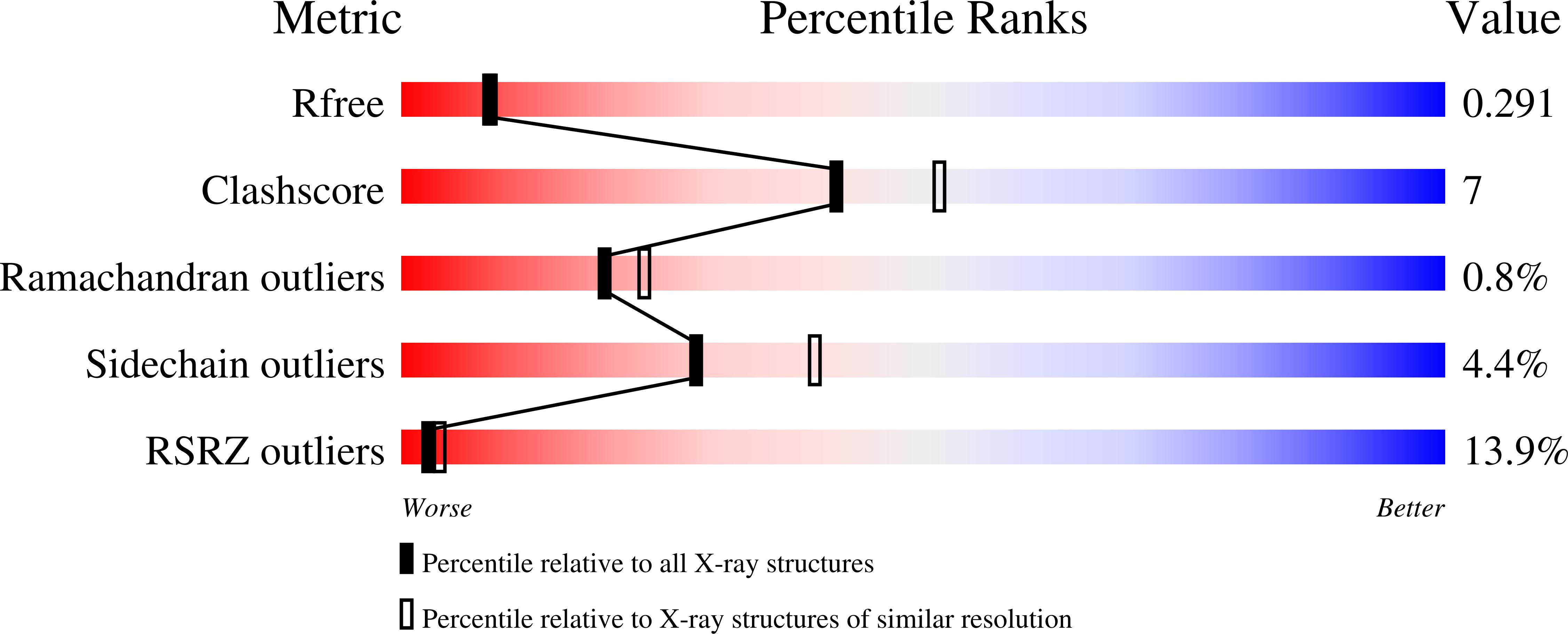
R-Value Free:
0.28
R-Value Work:
0.21
Space Group:
H 3 2