9EZD image
Deposition Date 2024-04-11
Release Date 2025-04-02
Last Version Date 2025-04-02
Entry Detail
PDB ID:
9EZD
Keywords:
Title:
BsmI (Bottom Nicking mutant) crystallized with Mg2+ and cognate dsDNA (Post-reactive complex)
Biological Source:
Source Organism:
Host Organism:
Method Details:
Experimental Method:
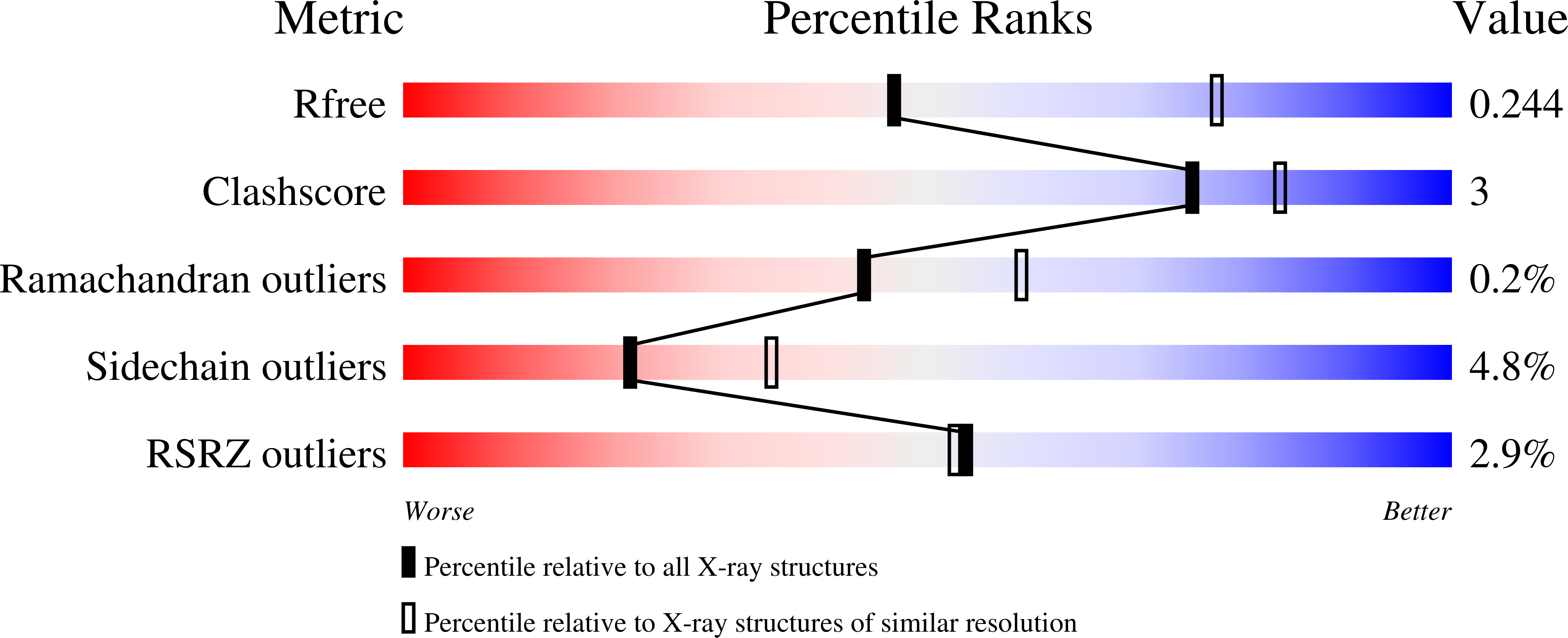
Resolution:
2.64 Å
R-Value Free:
0.25
R-Value Work:
0.21
R-Value Observed:
0.22
Space Group:
P 21 21 2
Macromolecular Entities
Polymer Type:polypeptide(L)
Molecule:BsmI
Gene (Uniprot):bsmIR
Mutations:R507D, G509V, E546V
Chain IDs:A
Chain Length:676
Number of Molecules:1
Biological Source:Geobacillus stearothermophilus
Polymer Type:polydeoxyribonucleotide
Molecule:DNA (Bottom strand - 5'-part)
Chain IDs:B (auth: E)
Chain Length:8
Number of Molecules:1
Biological Source:Geobacillus stearothermophilus
Polymer Type:polydeoxyribonucleotide
Molecule:DNA (Top strand)
Chain IDs:C (auth: F)
Chain Length:13
Number of Molecules:1
Biological Source:Geobacillus stearothermophilus
Polymer Type:polydeoxyribonucleotide
Molecule:DNA (Bottom strand - 3'-part)
Chain IDs:D (auth: G)
Chain Length:5
Number of Molecules:1
Biological Source:Geobacillus stearothermophilus
Primary Citation
Crystal structures of monomeric BsmI restriction endonuclease reveal coordinated sequential cleavage of two DNA strands.
Commun Biol 8 387 387 (2025)
PMID: 40055548 DOI: 10.1038/s42003-025-07612-z

Abstact

BsmI, a thermophilic Type IIS restriction endonuclease from Bacillus stearothermophilus, presents a unique structural composition, housing two distinct active sites within a single monomer. Recognition of the non-symmetrical 5'-GAATGC-3' sequence enables precise cleavage of the top and bottom DNA strands. Synthetic biology interventions have led to the transformation of BsmI into Nb.BsmI, a nicking endonuclease. Here we introduce Nt*.BsmI, tailored for top-strand cleavage, which is inactive on standard double-stranded DNA, but active on bottom-strand nicked DNA, suggesting a sequential cleavage mechanism. Crystallographic structures of pre- and post-reactive complexes with cognate DNA show one major conformational change, a retractable loop possibly governing sequential active site accessibility. The x-ray structures reveal the position of the divalent metal ions in the active sites and the DNA:protein interactions, while the models predicted by Alphafold3 are incorrect. This comprehensive structural and functional study lays a foundation for rational enzyme redesign and potential applications in biotechnology.

Legend

Protein

Chemical

Disease

Primary Citation of related structures