Deposition Date
2024-03-12
Release Date
2025-08-20
Last Version Date
2025-12-31
Entry Detail
PDB ID:
9EN4
Keywords:
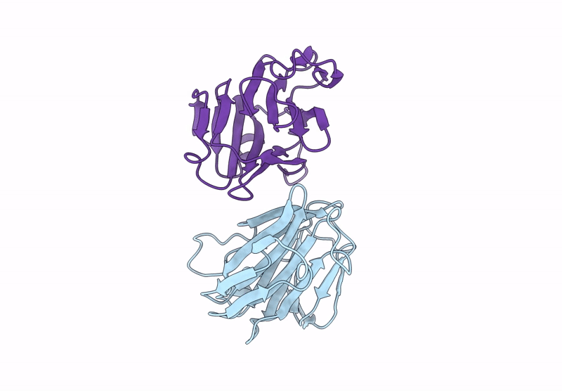
Title:
VP8* domain of the spike protein VP4 from bovine P[3] strain of rotavirus species C
Biological Source:
Source Organism(s):
Bovine rotavirus C (Taxon ID: 31588)
Expression System(s):
Method Details:
Experimental Method:
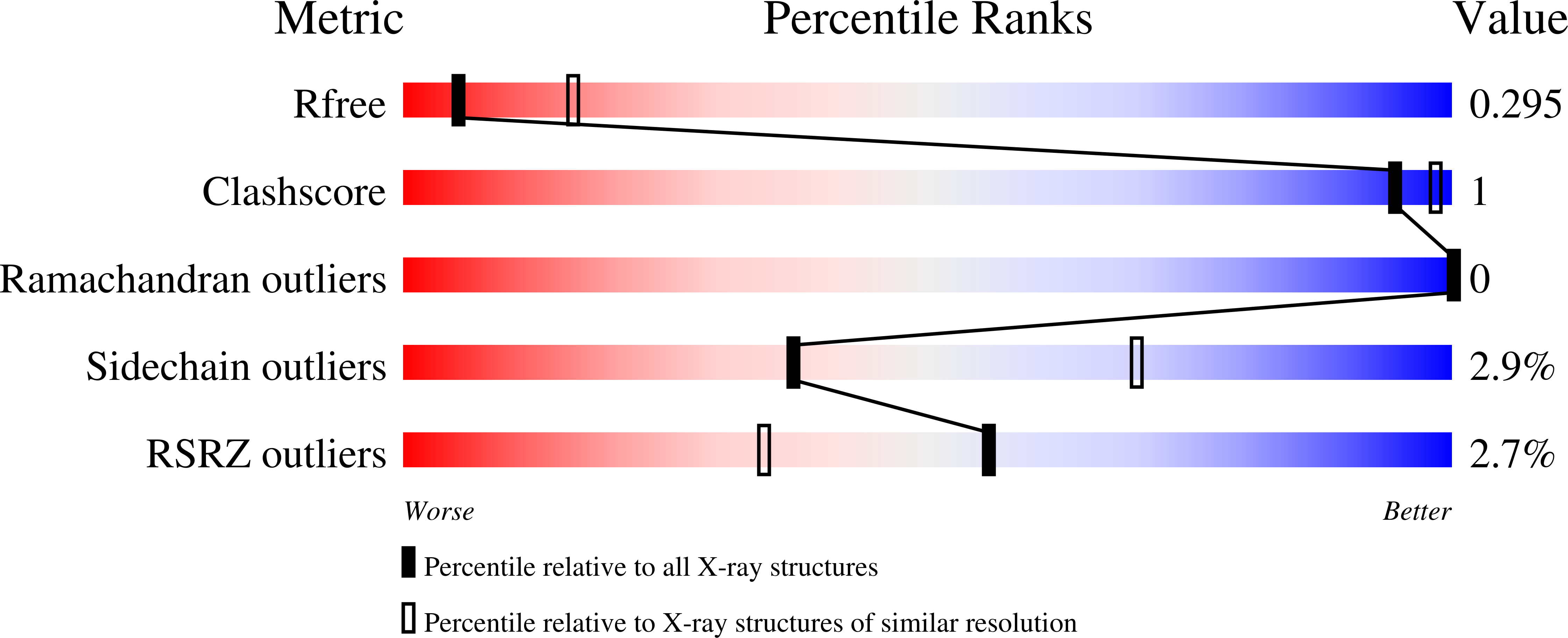
Resolution:
3.00 Å
R-Value Free:
0.29
R-Value Work:
0.24
R-Value Observed:
0.24
Space Group:
P 21 21 21