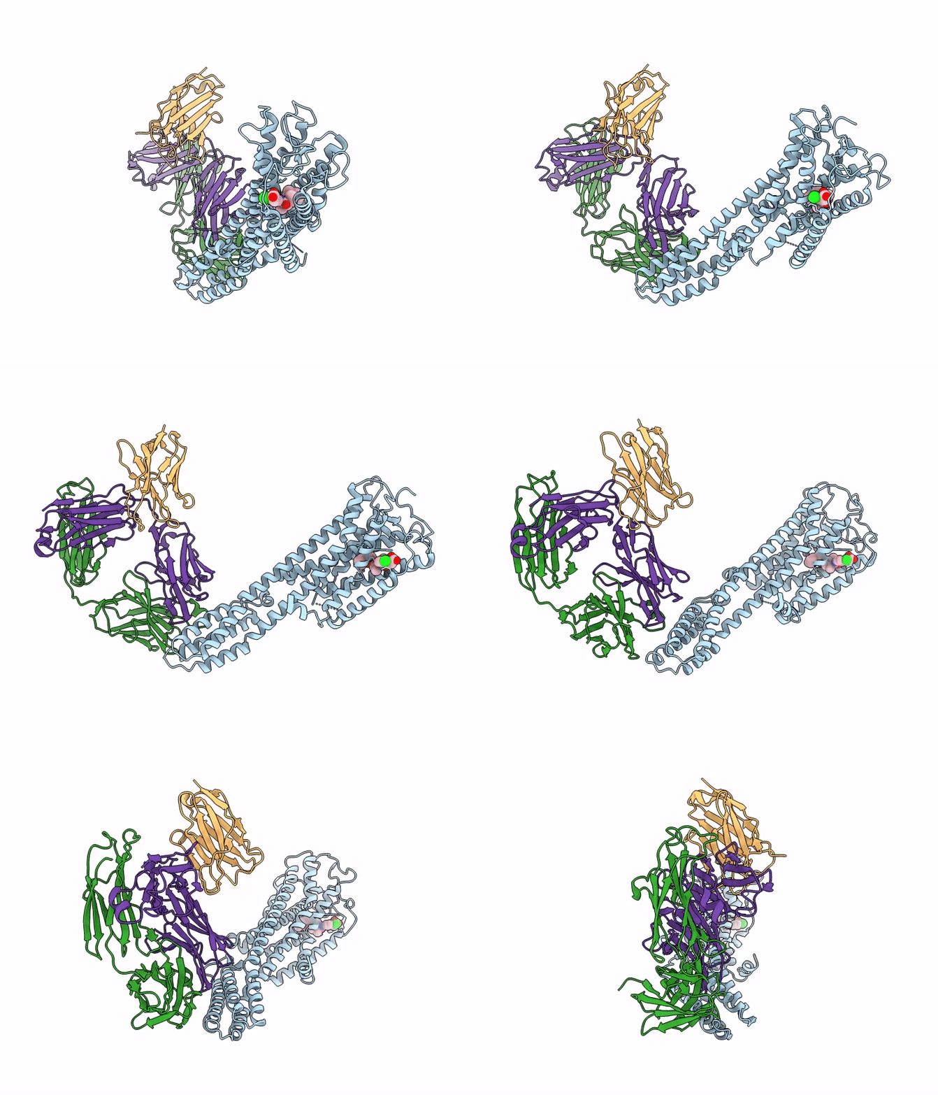
9EE5 image
Deposition Date 2024-11-18
Release Date 2025-09-03
Last Version Date 2025-10-22
Entry Detail
PDB ID:
9EE5
Title:
Cryo-EM structure of the ONO2550289-bound prostaglandin D2 receptor (DP1)-bRIL-Fab complex
Biological Source:
Source Organism:
Host Organism:
Method Details:
Experimental Method:
Resolution:
2.89 Å
Aggregation State:
PARTICLE
Reconstruction Method:
SINGLE PARTICLE
Macromolecular Entities
Protein Blast
Polymer Type:polypeptide(L)
Molecule:Prostaglandin D2 receptor,Soluble cytochrome b562
Gene (Uniprot):cybC, PTGDR
Chain IDs:D (auth: A)
Chain Length:495
Number of Molecules:1
Biological Source:Homo sapiens
Polymer Type:polypeptide(L)
Molecule:anti-BRIL Fab Heavy Chain
Chain IDs:A (auth: H)
Chain Length:229
Number of Molecules:1
Biological Source:synthetic construct
Polymer Type:polypeptide(L)
Molecule:anti-Fab Nanobody synthetic construct
Chain IDs:B (auth: K)
Chain Length:123
Number of Molecules:1
Biological Source:synthetic construct
Polymer Type:polypeptide(L)
Molecule:anti-BRIL Fab Light Chain
Chain IDs:C (auth: L)
Chain Length:214
Number of Molecules:1
Biological Source:synthetic construct
Ligand Molecules
Primary Citation
Structural insights into the mechanism of activation and inhibition of the prostaglandin D2 receptor 1.
Nat Commun 16 8944 8944 (2025)
PMID: 41062467 DOI: 10.1038/s41467-025-64002-z

Abstact

The prostaglandin D2 receptor 1 (DP1), a member of the prostanoid G protein-coupled receptor (GPCR) family, plays critical roles in allergic responses, sleep regulation, immune modulation, and vasodilation. Here, we present five high-resolution cryo-electron microscopy (cryo-EM) structures of the human DP1 receptor, including an apo structure, two inactive state structures bound to two different inverse agonists developed by ONO Pharmaceutical, and two active state structures in complex with the Gs protein and bound to the endogenous agonist PGD2 and its selective derivative BW245C. Structural analysis, complemented by molecular dynamics simulations and site-directed mutagenesis, reveals key residues involved in ligand recognition and suggests a distinct activation mechanism for DP1, which lacks most of the conserved class A GPCR motifs. Notably, the unique residue K76 within the conserved sodium pocket acts as a major activation switch, while amphiphilic helix 8 adopts an unconventional orientation essential for receptor function. These findings offer valuable insights into the structure and function of prostanoid receptors and may facilitate the development of therapeutics targeting DP1.

Legend

Protein

Chemical

Disease

Primary Citation of related structures