9EBX image
Entry Detail
PDB ID:
9EBX
Title:
Chimeric fluorescence biosensor formed from a lactate-binding protein and GFP
Biological Source:
Host Organism:
PDB Version:
Deposition Date:
2024-11-13
Release Date:
2025-03-19
Method Details:
Experimental Method:
Resolution:
2.42 Å
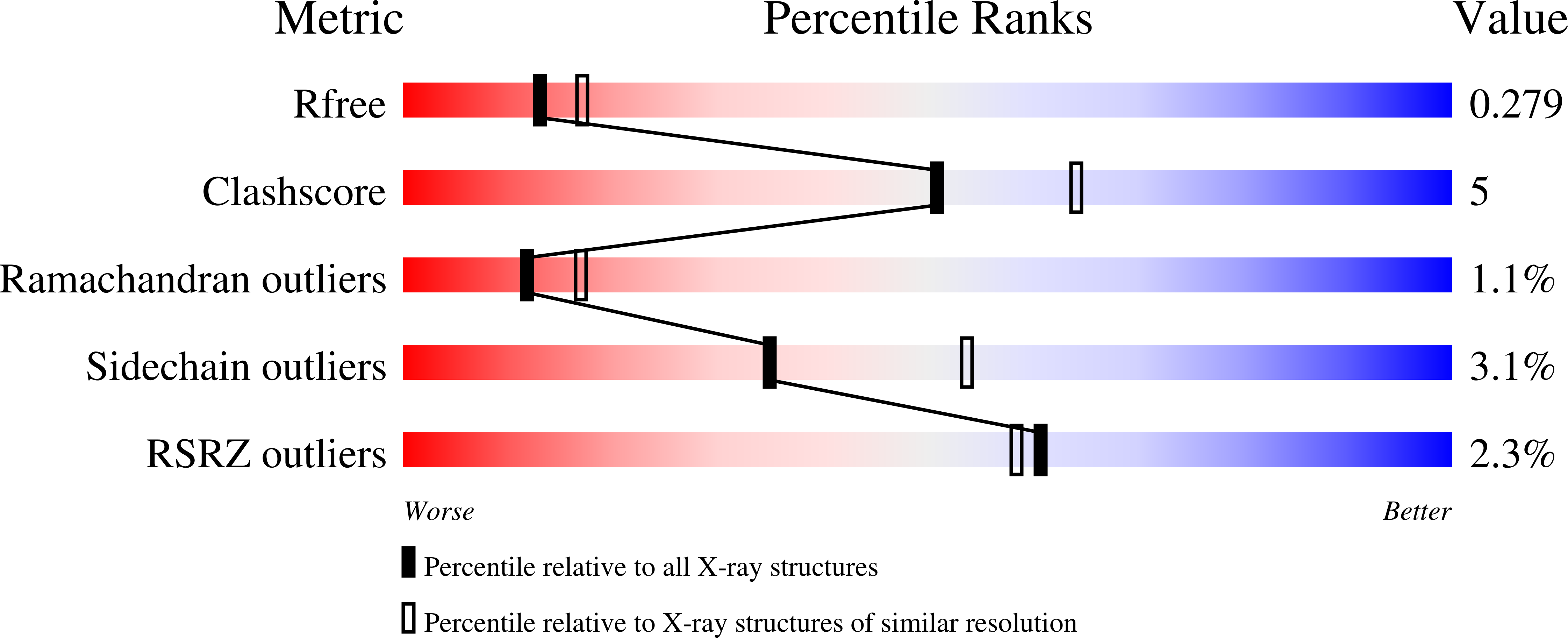
R-Value Free:
0.27
R-Value Work:
0.23
R-Value Observed:
0.23
Space Group:
P 1
Macromolecular Entities
Polymer Type:polypeptide(L)
Description:Green fluorescent protein,Methyl-accepting chemotaxis transducer (TlpC)
Chain IDs:A
Chain Length:517
Number of Molecules:1
Biological Source:Aequorea victoria, Helicobacter pylori
Modified Residue
Compound ID Chain ID Parent Comp ID Details 2D Image
SWG A SER chromophore
Primary Citation
State-dependent motion of a genetically encoded fluorescent biosensor.
Proc.Natl.Acad.Sci.USA 122 e2426324122 e2426324122 (2025)
PMID: 40048274 DOI: 10.1073/pnas.2426324122

Abstact

Genetically encoded biosensors can measure biochemical properties such as small-molecule concentrations with single-cell resolution, even in vivo. Despite their utility, these sensors are "black boxes": Very little is known about the structures of their low- and high-fluorescence states or what features are required to transition between them. We used LiLac, a lactate biosensor with a quantitative fluorescence-lifetime readout, as a model system to address these questions. X-ray crystal structures and engineered high-affinity metal bridges demonstrate that LiLac exhibits a large interdomain twist motion that pulls the fluorescent protein away from a "sealed," high-lifetime state in the absence of lactate to a "cracked," low-lifetime state in its presence. Understanding the structures and dynamics of LiLac will help to think about and engineer other fluorescent biosensors.

Legend

Protein

Chemical

Disease

Primary Citation of related structures