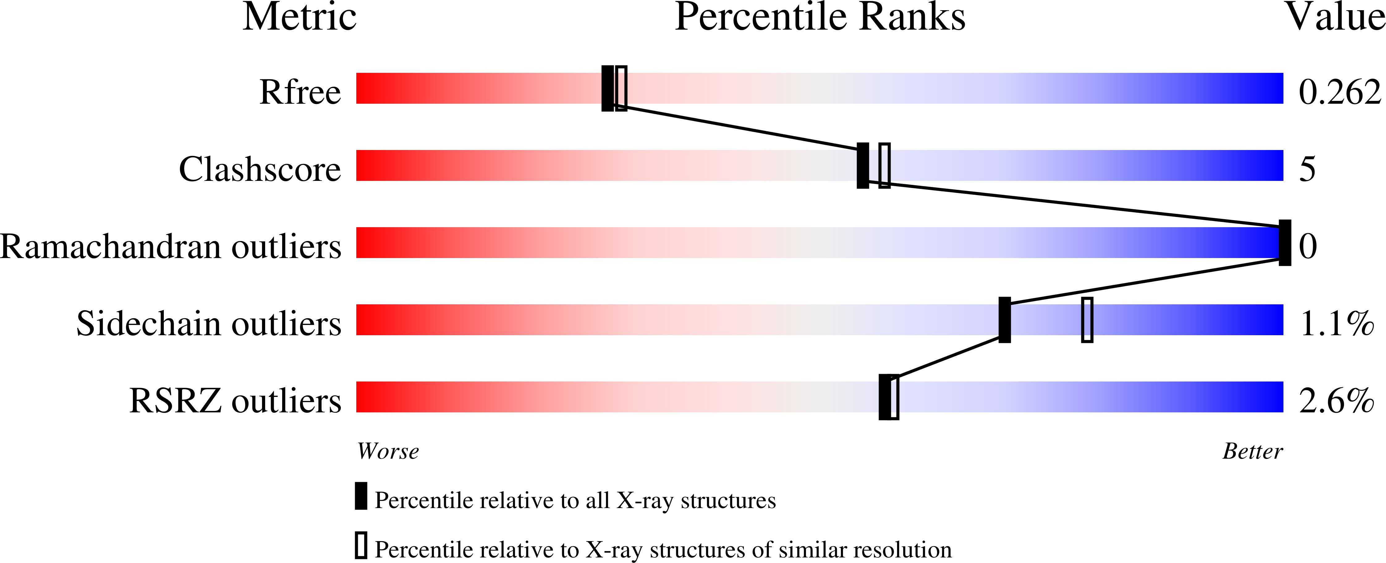
9EAW image
Entry Detail
PDB ID:
9EAW
Keywords:
Title:
Escherichia coli Carbonic Anhydrase 2 in Space Group P2(1)2(1)2
Biological Source:
PDB Version:
Deposition Date:
2024-11-11
Release Date:
2024-11-20
Method Details:
Experimental Method:
Resolution:
2.26 Å
R-Value Free:
0.26
R-Value Work:
0.19
R-Value Observed:
0.20
Space Group:
P 21 21 2
Macromolecular Entities
Polymer Type:polypeptide(L)
Description:Carbonic anhydrase
Chain IDs:A, B
Chain Length:220
Number of Molecules:2
Biological Source:Escherichia coli BL21(DE3)
Ligand Molecules
Primary Citation
Serendipitous high-resolution structure of Escherichia coli carbonic anhydrase 2.
Acta Crystallogr.,Sect.F 81 47 52 (2025)
PMID: 39812168 DOI: 10.1107/S2053230X25000068

Abstact

X-ray crystallography remains the dominant method of determining the three-dimensional structure of proteins. Nevertheless, this resource-intensive process may be hindered by the unintended crystallization of contaminant proteins from the expression source. Here, the serendipitous discovery of two novel crystal forms and one new, high-resolution structure of carbonic anhydrase 2 (CA2) from Escherichia coli that arose during a crystallization campaign for an unrelated target is reported. By comparing unit-cell parameters with those in the PDB, contaminants such as CA2 can be identified, preventing futile molecular-replacement attempts. Crystallographers can use these new lattice parameters to diagnose CA2 contamination in similar experiments.

Legend

Protein

Chemical

Disease

Primary Citation of related structures