Deposition Date
2024-11-04
Release Date
2025-01-29
Last Version Date
2025-05-14
Entry Detail
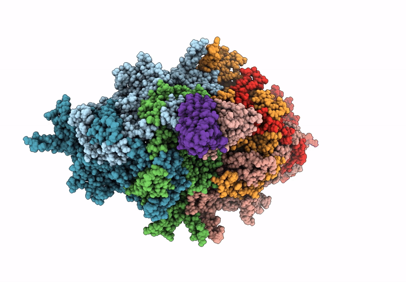
PDB ID:
9E7X
Keywords:
Title:
Canine parvovirus subtype 2a empty capsid in complex with Fab fragments of Mab 2C5
Biological Source:
Source Organism(s):
Canine parvovirus 2a (Taxon ID: 497961)
Canis lupus familiaris (Taxon ID: 9615)
Canis lupus familiaris (Taxon ID: 9615)
Expression System(s):
Method Details:
Experimental Method:
Resolution:
3.20 Å
Aggregation State:
PARTICLE
Reconstruction Method:
SINGLE PARTICLE