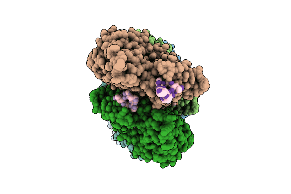
9DUU image
Entry Detail
PDB ID:
9DUU
EMDB ID:
Title:
Cryo-EM structure of recombinant wildtype ACTA1 phalloidin-stabilized F-actin
Biological Source:
Host Organism:
PDB Version:
Deposition Date:
2024-10-04
Release Date:
2024-10-23
Method Details:
Experimental Method:
Resolution:
3.40 Å
Aggregation State:
FILAMENT
Reconstruction Method:
HELICAL
Macromolecular Entities
Polymer Type:polypeptide(L)
Description:Actin, alpha skeletal muscle
Chain IDs:A, B, C, D, E, F
Chain Length:375
Number of Molecules:6
Biological Source:Homo sapiens
Polymer Type:polypeptide(L)
Description:phalloidin
Chain IDs:G (auth: M), H (auth: N), I (auth: O), J (auth: P), K (auth: Q), L (auth: R), M (auth: S)
Chain Length:7
Number of Molecules:7
Biological Source:Amanita phalloides
Modified Residue
Compound ID Chain ID Parent Comp ID Details 2D Image
HIC A HIS modified residue
Primary Citation
Dilated cardiomyopathy-associated skeletal muscle actin (ACTA1) mutation R256H disrupts actin structure and function and causes cardiomyocyte hypocontractility.
Proc.Natl.Acad.Sci.USA 121 e2405020121 e2405020121 (2024)
PMID: 39503885 DOI: 10.1073/pnas.2405020121

Abstact

Skeletal muscle actin (ACTA1) mutations are a prevalent cause of skeletal myopathies consistent with ACTA1's high expression in skeletal muscle. Rare de novo mutations in ACTA1 associated with combined cardiac and skeletal myopathies have been reported, but ACTA1 represents only ~20% of the total actin pool in cardiomyocytes, making its role in cardiomyopathy controversial. Here we demonstrate how a mutation in an actin isoform expressed at low levels in cardiomyocytes can cause cardiomyopathy by focusing on a unique ACTA1 variant, R256H. We previously identified this variant in a family with dilated cardiomyopathy, who had reduced systolic function without clinical skeletal myopathy. Using a battery of multiscale biophysical tools, we show that R256H has potent effects on ACTA1 function at the molecular scale and in human cardiomyocytes. Importantly, we demonstrate that R256H acts in a dominant manner, where the incorporation of small amounts of mutant protein into thin filaments is sufficient to disrupt molecular contractility, and that this effect is dependent on the presence of troponin and tropomyosin. To understand the structural basis of this change in regulation, we resolved a structure of R256H filaments using cryoelectron microscopy, and we see alterations in actin's structure that have the potential to disrupt interactions with tropomyosin. Finally, we show that ACTA1R256H/+ human-induced pluripotent stem cell cardiomyocytes demonstrate reduced contractility and sarcomeric organization. Taken together, we demonstrate that R256H has multiple effects on ACTA1 function that are sufficient to cause reduced contractility and establish a likely causative relationship between ACTA1 R256H and clinical cardiomyopathy.

Legend

Protein

Chemical

Disease

Primary Citation of related structures