9DRV image
Deposition Date 2024-09-26
Release Date 2025-02-12
Last Version Date 2025-02-19
Entry Detail
PDB ID:
9DRV
Keywords:
Title:
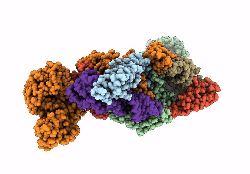
Crystal structure of M. tuberculosis PheRS-tRNA complex bound to inhibitor D-004
Biological Source:
Source Organism:
Method Details:
Experimental Method:
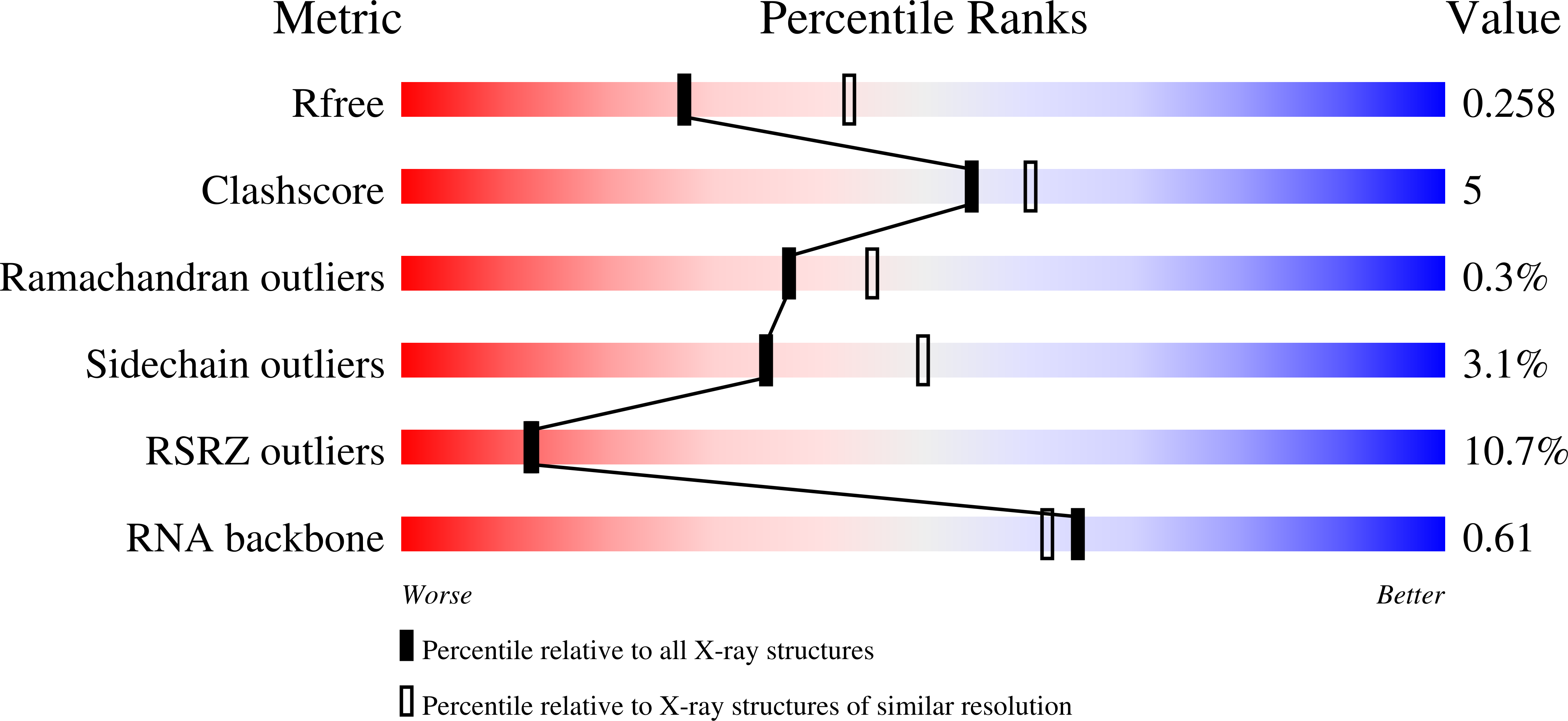
Resolution:
2.46 Å
R-Value Free:
0.25
R-Value Work:
0.20
R-Value Observed:
0.21
Space Group:
P 1 21 1
Macromolecular Entities
Polymer Type:polypeptide(L)
Molecule:Phenylalanine--tRNA ligase alpha subunit
Gene (Uniprot):pheS
Chain IDs:A, D
Chain Length:350
Number of Molecules:2
Biological Source:Mycobacterium tuberculosis H37Rv
Polymer Type:polypeptide(L)
Molecule:Phenylalanine--tRNA ligase beta subunit
Gene (Uniprot):pheT
Chain IDs:B, E
Chain Length:835
Number of Molecules:2
Biological Source:Mycobacterium tuberculosis H37Rv
Polymer Type:polyribonucleotide
Molecule:tRNA(phe)
Chain IDs:C, F
Chain Length:77
Number of Molecules:2
Biological Source:Mycobacterium tuberculosis (strain ATCC 25618 / H37Rv)
Primary Citation
Different chemical scaffolds bind to L-phe site in Mycobacterium tuberculosis Phe-tRNA synthetase.
Eur.J.Med.Chem. 287 117335 117335 (2025)
PMID: 39919438 DOI: 10.1016/j.ejmech.2025.117335

Abstact

Tuberculosis (TB), caused by Mycobacterium tuberculosis (Mt), is one of the deadliest infectious diseases. The rise of multidrug-resistant strains represents a major public health threat, requiring new therapeutic options. Bacterial aminoacyl-tRNA synthetases (aaRS) have been shown to be highly promising drug targets, including for TB treatment. These enzymes play an essential role in translating the DNA gene code into protein sequence by attaching specific amino acid to their cognate tRNAs. They have multiple binding sites that can be targeted for inhibitor discovery: amino acid binding pocket, ATP binding pocket, tRNA binding site and an editing domain. Recently we reported several high-resolution structures of M. tuberculosis phenylalanyl-tRNA synthetase (MtPheRS) complexed with tRNAPhe and either L-Phe or a nonhydrolyzable phenylalanine adenylate analog. Here, using Nucleic Magnetic Resonance (NMR) and Surface Plasmon Resonance (SPR) we identified fragments that bind to MtPheRS and we determined crystal structures of their complexes with MtPheRS/tRNAPhe. All the binders interact with the L-Phe amino acid binding site. The analysis of interactions of the new compounds combined with adenylate analog structure provides insights for the rational design of anti-tuberculosis drugs. The 3' arm of the tRNAPhe in all the structures was disordered with exception of one complex with D-735 compound. In this structure the 3' CCA end of the acceptor stem is observed in the editing domain of MtPheRS providing insights regarding the post-transfer editing activity of class II aaRS.

Legend

Protein

Chemical

Disease

Primary Citation of related structures