
Deposition Date
2024-09-03
Release Date
2025-05-14
Last Version Date
2025-05-14
Entry Detail
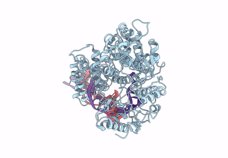
PDB ID:
9DGY
Keywords:
Title:
Mycobacterium tuberculosis UvrD1 monomer-DNA complex
Biological Source:
Source Organism(s):
Mycobacterium tuberculosis (Taxon ID: 1773)
Expression System(s):
Method Details:
Experimental Method:
Resolution:
7.00 Å
Aggregation State:
PARTICLE
Reconstruction Method:
SINGLE PARTICLE
