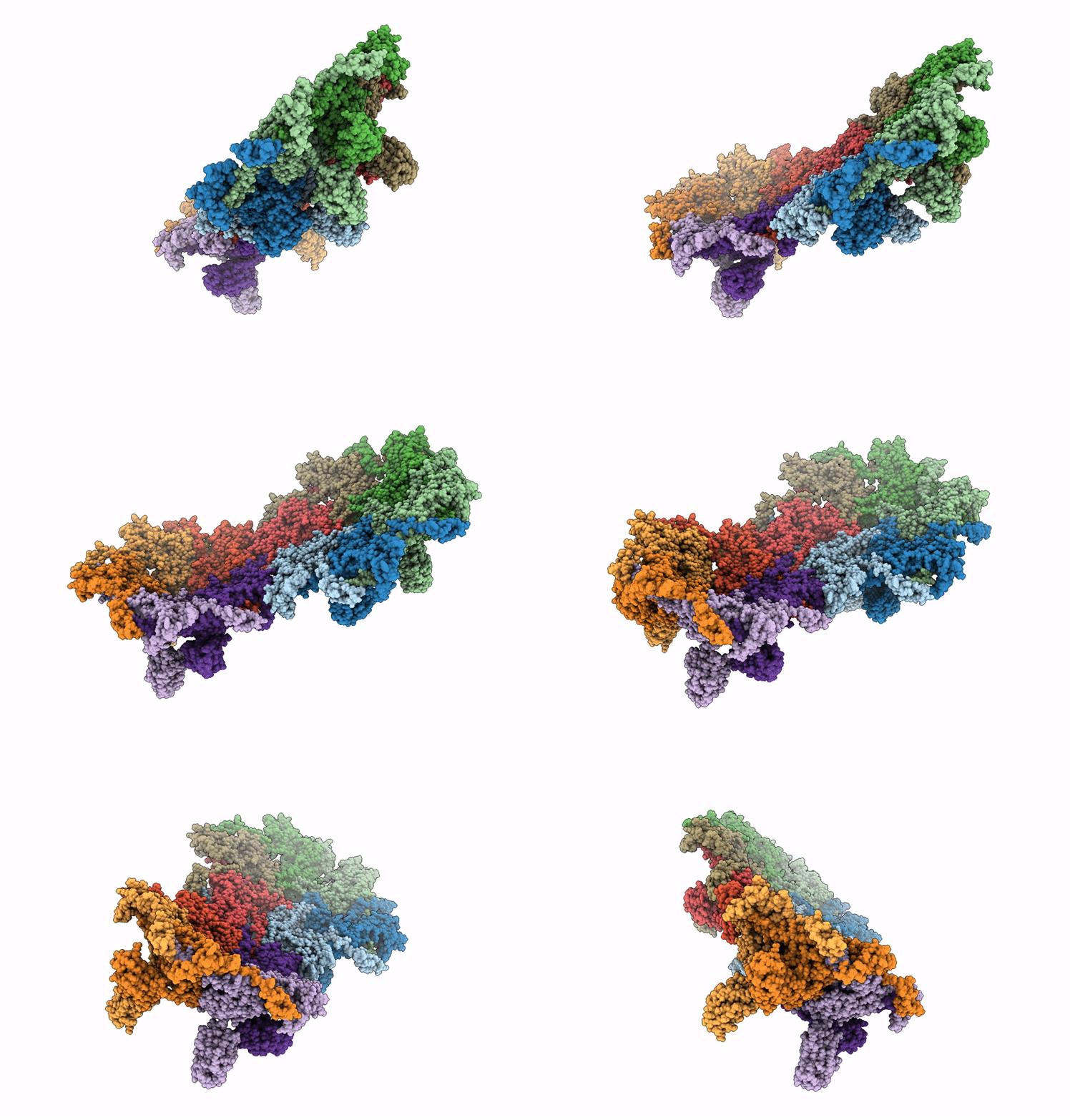
9D9X image
Deposition Date 2024-08-21
Release Date 2024-09-25
Last Version Date 2025-06-11
Entry Detail
PDB ID:
9D9X
Keywords:
Title:
Mycobacteriophage Bxb1 Capsid - Composite map and model
Biological Source:
Source Organism:
Method Details:
Experimental Method:
Resolution:
3.00 Å
Aggregation State:
PARTICLE
Reconstruction Method:
SINGLE PARTICLE
Macromolecular Entities
Polymer Type:polypeptide(L)
Molecule:Major capsid protein
Gene (Uniprot):14
Chain IDs:A (auth: Ha), B (auth: Hb), C (auth: Hc), D (auth: Hd), E (auth: He), F (auth: Hf), G (auth: Pa), H (auth: Pb), I (auth: Pc), J (auth: Pd), K (auth: Pe)
Chain Length:397
Number of Molecules:11
Biological Source:Mycobacterium phage Bxb1
Ligand Molecules
Primary Citation
Structure and infection dynamics of mycobacteriophage Bxb1.
Cell 188 2925 ? (2025)
PMID: 40239650 DOI: 10.1016/j.cell.2025.03.027

Abstact

Mycobacteriophage Bxb1 is a well-characterized virus of Mycobacterium smegmatis with double-stranded DNA and a long, flexible tail. Mycobacteriophages show considerable potential as therapies for Mycobacterium infections, but little is known about the structural details of these phages or how they bind to and traverse the complex Mycobacterium cell wall. Here, we report the complete structure and atomic model of phage Bxb1, including the arrangement of immunodominant domains of both the capsid and tail tube subunits, as well as the assembly of the protein subunits in the tail-tip complex. The structure contains protein assemblies with 3-, 5-, 6-, and 12-fold symmetries, which interact to satisfy several symmetry mismatches. Cryoelectron tomography of phage particles bound to M. smegmatis reveals the structural transitions that occur for free phage particles to bind to the cell surface and navigate through the cell wall to enable DNA transfer into the cytoplasm.

Legend

Protein

Chemical

Disease

Primary Citation of related structures