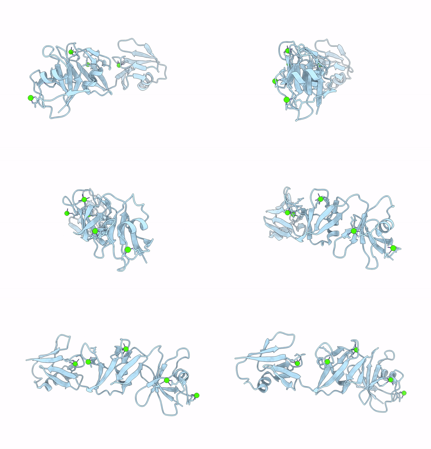
9D5A image
Entry Detail
PDB ID:
9D5A
EMDB ID:
Keywords:
Title:
Structure of Citrobacter multi-ubiquitin protein, local refinement of one full-length protomer
Biological Source:
PDB Version:
Deposition Date:
2024-08-13
Release Date:
2025-04-02
Method Details:
Experimental Method:
Resolution:
2.73 Å
Aggregation State:
FILAMENT
Reconstruction Method:
SINGLE PARTICLE
Macromolecular Entities
Polymer Type:polypeptide(L)
Description:Multi-ubiquitin domain-containing protein
Chain IDs:A
Chain Length:247
Number of Molecules:1
Biological Source:Citrobacter sp. RHBSTW-00271
Ligand Molecules
Primary Citation
Structural diversity and oligomerization of bacterial ubiquitin-like proteins.
Structure ? ? ? (2025)
PMID: 40250427 DOI: 10.1016/j.str.2025.03.011

Abstact

Bacteria possess a variety of operons with homology to eukaryotic ubiquitination pathways that encode predicted E1, E2, E3, deubiquitinase, and ubiquitin-like proteins. Some of these pathways have recently been shown to function in anti-bacteriophage immunity, but the biological functions of others remain unknown. Here, we show that ubiquitin-like proteins in two bacterial operon families show surprising architectural diversity, possessing one to three β-grasp domains preceded by diverse N-terminal domains. We find that a large group of bacterial ubiquitin-like proteins possess three β-grasp domains and form homodimers and helical filaments mediated by conserved Ca2+ ion binding sites. Our findings highlight a distinctive mode of self-assembly for ubiquitin-like proteins and suggest that Ca2+-mediated ubiquitin-like protein filament assembly and/or disassembly enables cells to sense and respond to stress conditions that alter intracellular metal ion concentration.

Legend

Protein

Chemical

Disease

Primary Citation of related structures