9D58 image
Deposition Date 2024-08-13
Release Date 2025-03-12
Last Version Date 2025-03-12
Entry Detail
PDB ID:
9D58
Title:
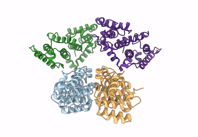
Human Dystrophin tandem calponin homology actin-binding domain crystallized in a closed-state conformation
Biological Source:
Source Organism:
Homo sapiens (Taxon ID: 9606)
Host Organism:
Method Details:
Experimental Method:
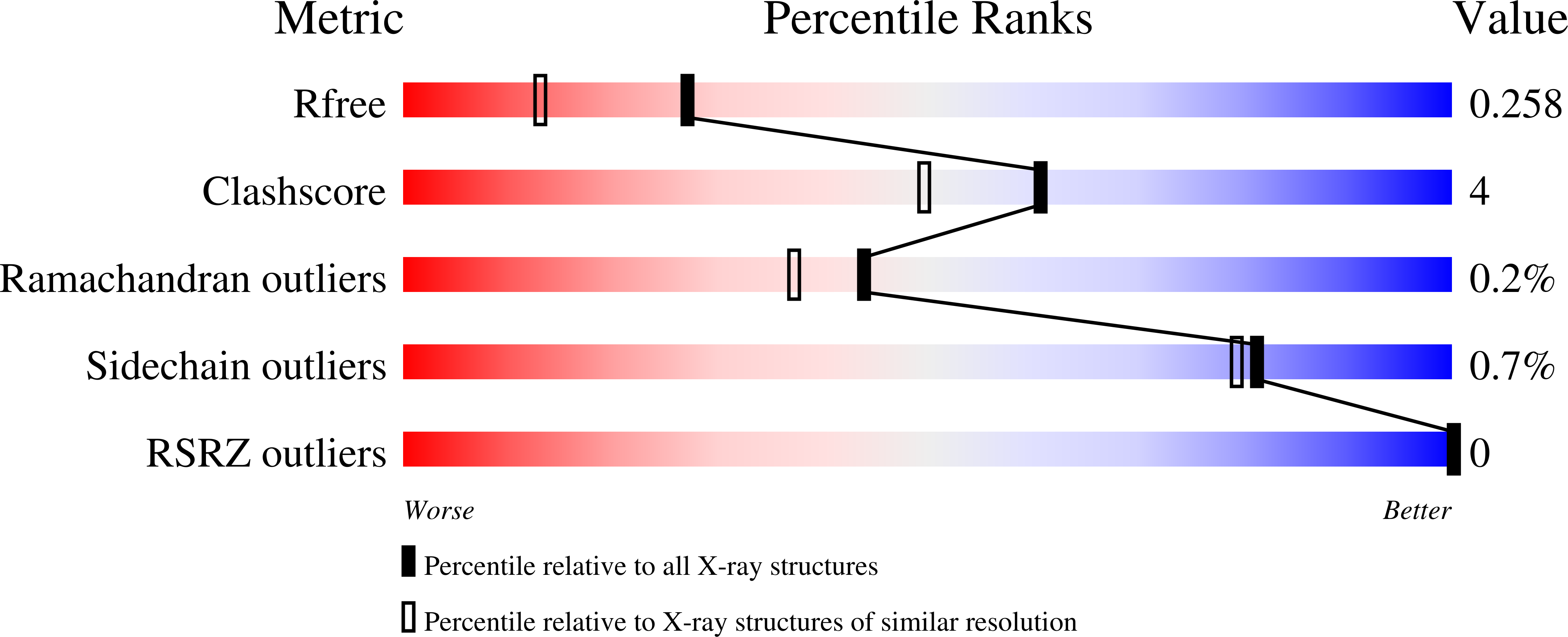
Resolution:
1.94 Å
R-Value Free:
0.26
R-Value Work:
0.21
R-Value Observed:
0.24
Space Group:
P 1 21 1
Macromolecular Entities
Polymer Type:polypeptide(L)
Molecule:Dystrophin
Gene (Uniprot):DMD
Chain IDs:A, B, C, D
Chain Length:248
Number of Molecules:4
Biological Source:Homo sapiens
Primary Citation
Human dystrophin tandem calponin homology actin-binding domain crystallized in a closed-state conformation.
Acta Crystallogr D Struct Biol 81 122 129 (2025)
PMID: 40007458 DOI: 10.1107/S2059798325001457

Abstact

The structure of the N-terminal actin-binding domain of human dystrophin was determined at 1.94 Å resolution. Each chain in the asymmetric unit exists in a `closed' conformation, with the first and second calponin homology (CH) domains directly interacting via a 2500.6 Å2 interface. The positioning of the individual CH domains is comparable to the domain-swapped dimer seen in previous human dystrophin and utrophin actin-binding domain 1 structures. The CH1 domain is highly similar to the actin-bound utrophin structure and structural homology suggests that the `closed' single-chain conformation opens during actin binding to mitigate steric clashes between CH2 and actin.

Legend

Protein

Chemical

Disease

Primary Citation of related structures