Deposition Date
2024-08-11
Release Date
2025-08-13
Last Version Date
2025-10-15
Entry Detail
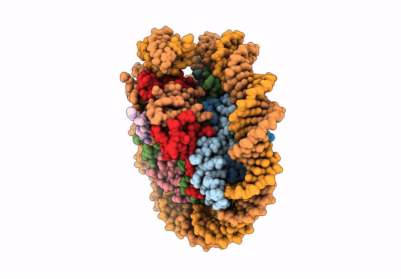
PDB ID:
9D3K
Keywords:
Title:
Two Dsup molecules in complex with the nucleosome open from both sides
Biological Source:
Source Organism(s):
Homo sapiens (Taxon ID: 9606)
Ramazzottius varieornatus (Taxon ID: 947166)
synthetic construct (Taxon ID: 32630)
Ramazzottius varieornatus (Taxon ID: 947166)
synthetic construct (Taxon ID: 32630)
Expression System(s):
Method Details:
Experimental Method:
Resolution:
2.70 Å
Aggregation State:
PARTICLE
Reconstruction Method:
SINGLE PARTICLE