9D2B image
Deposition Date 2024-08-08
Release Date 2025-08-13
Last Version Date 2025-11-12
Entry Detail
PDB ID:
9D2B
Keywords:
Title:
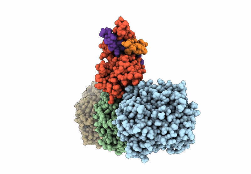
Symmetry-expanded reconstruction of augmin T-II bonsai on the microtubule
Biological Source:
Source Organism:
Xenopus laevis (Taxon ID: 8355)
Bos taurus (Taxon ID: 9913)
Host Organism:
Method Details:
Experimental Method:
Resolution:
3.08 Å
Aggregation State:
FILAMENT
Reconstruction Method:
SINGLE PARTICLE
Macromolecular Entities
Structures with similar UniProt ID
Protein Blast
Polymer Type:polypeptide(L)
Molecule:Tubulin beta-2B chain
Gene (Uniprot):TUBB2B
Chain IDs:B
Chain Length:445
Number of Molecules:1
Biological Source:Bos taurus
Structures with similar UniProt ID
Protein Blast
Polymer Type:polypeptide(L)
Molecule:Tubulin alpha-1B chain
Chain IDs:A (auth: C), C (auth: A)
Chain Length:451
Number of Molecules:2
Biological Source:Bos taurus
Structures with similar UniProt ID
Protein Blast
Polymer Type:polypeptide(L)
Molecule:HAUS augmin like complex subunit 6 L homeolog isoform X1
Gene (Uniprot):haus6.L
Chain IDs:D (auth: F)
Chain Length:289
Number of Molecules:1
Biological Source:Xenopus laevis
Structures with similar UniProt ID
Protein Blast
Polymer Type:polypeptide(L)
Molecule:HAUS augmin like complex subunit 7 S homeolog
Gene (Uniprot):haus7.S
Chain IDs:E (auth: G)
Chain Length:282
Number of Molecules:1
Biological Source:Xenopus laevis
Structures with similar UniProt ID
Protein Blast
Polymer Type:polypeptide(L)
Molecule:HAUS augmin-like complex subunit 8
Gene (Uniprot):haus8
Chain IDs:F (auth: H)
Chain Length:260
Number of Molecules:1
Biological Source:Xenopus laevis
Primary Citation
How augmin establishes the angle of the microtubule branch site.
Nat Commun 16 9646 9646 (2025)
PMID: 41173848 DOI: 10.1038/s41467-025-64650-1

Abstact

How microtubules (MTs) are generated in the proper orientation is essential to understanding how the cytoskeleton organizes a cell and MT-dependent events such as cell division. In the spindle, most MTs are generated through the branching MT nucleation pathway. In this pathway, new MTs are nucleated from the side of existing MTs and oriented at a shallow angle by the branching factor augmin, ensuring that both MTs have the same polarity. Yet, how augmin binds MTs and sets the branch angle has remained unclear. Here, we report the cryo-electron microscopy structure of an augmin subcomplex on the MT. This structure resembles that of NDC80 bound to the MT, with the conserved CH domain of augmin's Haus6 subunit directly proximal to the MT lattice. We find that the Haus6 CH domain is a bona fide MT binding site that increases augmin's affinity for the MT and helps establish branch angle. A second binding site, located in the disordered N-terminus of Haus8, also establishes branch angle,. Thus, we find that augmin regulates MT branching using two domains, each tuned to modulate MT affinity and MT branch angle. This work expands our mechanistic understanding of branching MT nucleation and thus spindle formation.

Legend

Protein

Chemical

Disease

Primary Citation of related structures
Feedback Form
Name
Email
Institute
Feedback