9D0A image
Entry Detail
PDB ID:
9D0A
EMDB ID:
Keywords:
Title:
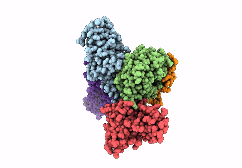
CryoEM structure of PAR2 with endogenous tethered ligand.
Biological Source:
Source Organism:
Host Organism:
PDB Version:
Deposition Date:
2024-08-06
Release Date:
2025-05-07
Method Details:
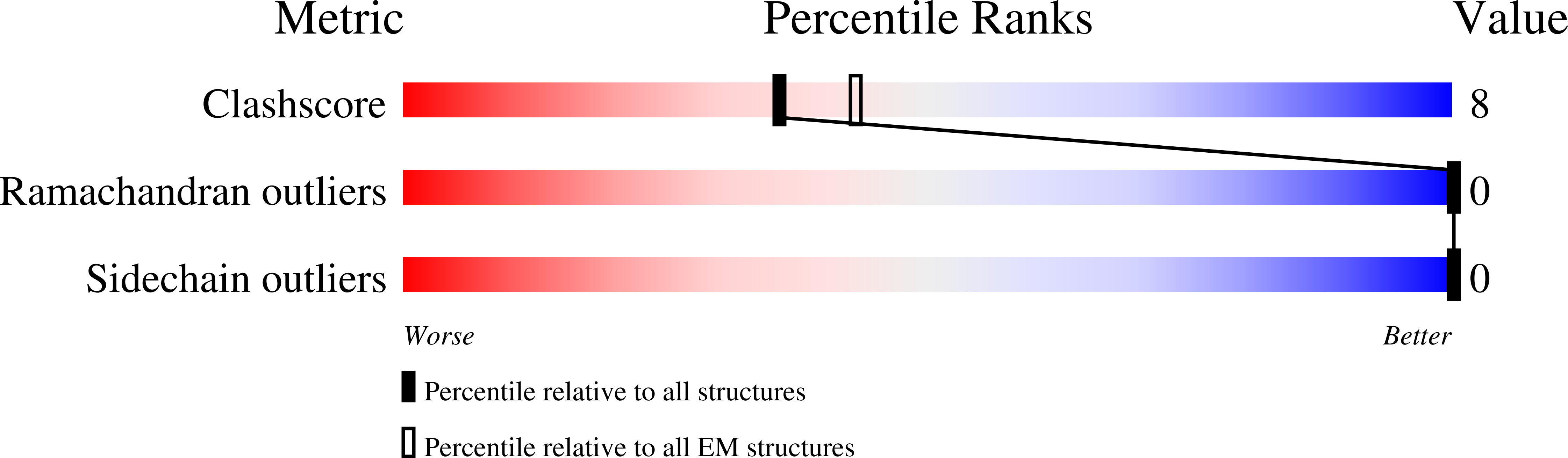
Experimental Method:
Resolution:
3.10 Å
Aggregation State:
PARTICLE
Reconstruction Method:
SINGLE PARTICLE
Macromolecular Entities
Polymer Type:polypeptide(L)
Description:Guanine nucleotide-binding protein G(q) subunit alpha, Guanine nucleotide-binding protein G(s) subunit alpha isoforms short chimera,Guanine nucleotide-binding protein G(s) subunit alpha isoforms short
Chain IDs:E (auth: A)
Chain Length:360
Number of Molecules:1
Biological Source:Homo sapiens
Polymer Type:polypeptide(L)
Description:Guanine nucleotide-binding protein G(I)/G(S)/G(T) subunit beta-1
Chain IDs:A (auth: B)
Chain Length:339
Number of Molecules:1
Biological Source:Homo sapiens
Polymer Type:polypeptide(L)
Description:scFv16
Chain IDs:C (auth: E)
Chain Length:251
Number of Molecules:1
Biological Source:unidentified
Polymer Type:polypeptide(L)
Description:Guanine nucleotide-binding protein G(I)/G(S)/G(O) subunit gamma-2
Chain IDs:B (auth: G)
Chain Length:59
Number of Molecules:1
Biological Source:Homo sapiens
Polymer Type:polypeptide(L)
Description:Proteinase-activated receptor 2
Chain IDs:D (auth: R)
Chain Length:397
Number of Molecules:1
Biological Source:Homo sapiens
Ligand Molecules
Primary Citation
Structural basis for the activation of proteinase-activated receptors PAR1 and PAR2.
Nat Commun 16 3931 3931 (2025)
PMID: 40287415 DOI: 10.1038/s41467-025-59138-x

Abstact

Members of the proteinase-activated receptor (PAR) subfamily of G protein-coupled receptors (GPCRs) play critical roles in processes like hemostasis, thrombosis, development, wound healing, inflammation, and cancer progression. Comprising PAR1-PAR4, these receptors are specifically activated by protease cleavage at their extracellular amino terminus, revealing a 'tethered ligand' that self-activates the receptor. This triggers complex intracellular signaling via G proteins and beta-arrestins, linking external protease signals to cellular functions. To date, direct structural visualization of these ligand-receptor complexes has been limited. Here, we present structural snapshots of activated PAR1 and PAR2 bound to their endogenous tethered ligands, revealing a shallow and constricted orthosteric binding pocket. Comparisons with antagonist-bound structures show minimal conformational changes in the TM6 helix and larger movements of TM7 upon activation. These findings reveal a common activation mechanism for PAR1 and PAR2, highlighting critical residues involved in ligand recognition. Additionally, the structure of PAR2 bound to a pathway selective antagonist, GB88, demonstrates how potent orthosteric engagement can be achieved by a small molecule mimicking the endogenous tethered ligand's interactions.

Legend

Protein

Chemical

Disease

Primary Citation of related structures