
Deposition Date
2024-08-02
Release Date
2024-12-18
Last Version Date
2024-12-18
Entry Detail
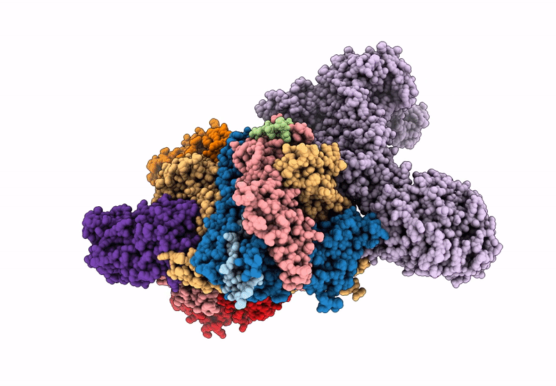
Biological Source:
Source Organism(s):
Mammalian orthoreovirus 3 Dearing (Taxon ID: 10886)
Method Details:
Experimental Method:
Resolution:
3.70 Å
Aggregation State:
PARTICLE
Reconstruction Method:
SINGLE PARTICLE
