
Deposition Date
2024-08-02
Release Date
2024-08-21
Last Version Date
2024-11-13
Entry Detail
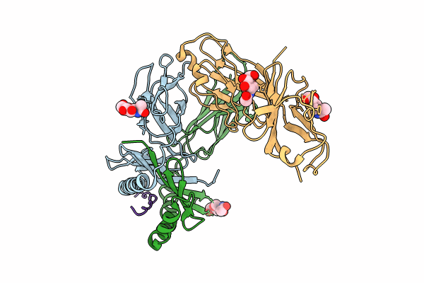
PDB ID:
9CYL
Keywords:
Title:
Structure of LAG3 loop1 deletion bound to the MHC class II molecule I-A(b)
Biological Source:
Source Organism(s):
Mus musculus (Taxon ID: 10090)
Homo sapiens (Taxon ID: 9606)
Homo sapiens (Taxon ID: 9606)
Expression System(s):
Method Details:
Experimental Method:
Resolution:
4.66 Å
R-Value Free:
0.32
R-Value Work:
0.26
R-Value Observed:
0.27
Space Group:
P 65 2 2
