
Deposition Date
2024-07-30
Release Date
2024-08-21
Last Version Date
2024-09-18
Entry Detail
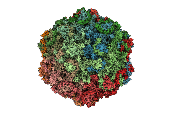
Biological Source:
Source Organism(s):
Bufavirus-1 (Taxon ID: 1209382)
Expression System(s):
Method Details:
Experimental Method:
Resolution:
2.73 Å
Aggregation State:
PARTICLE
Reconstruction Method:
SINGLE PARTICLE
