9CV8 image
Entry Detail
PDB ID:
9CV8
Keywords:
Title:
Crystal Structure of Cytochrome P450 NysL bound to nystatin
Biological Source:
Source Organism:
Host Organism:
PDB Version:
Deposition Date:
2024-07-28
Release Date:
2025-01-29
Method Details:
Experimental Method:
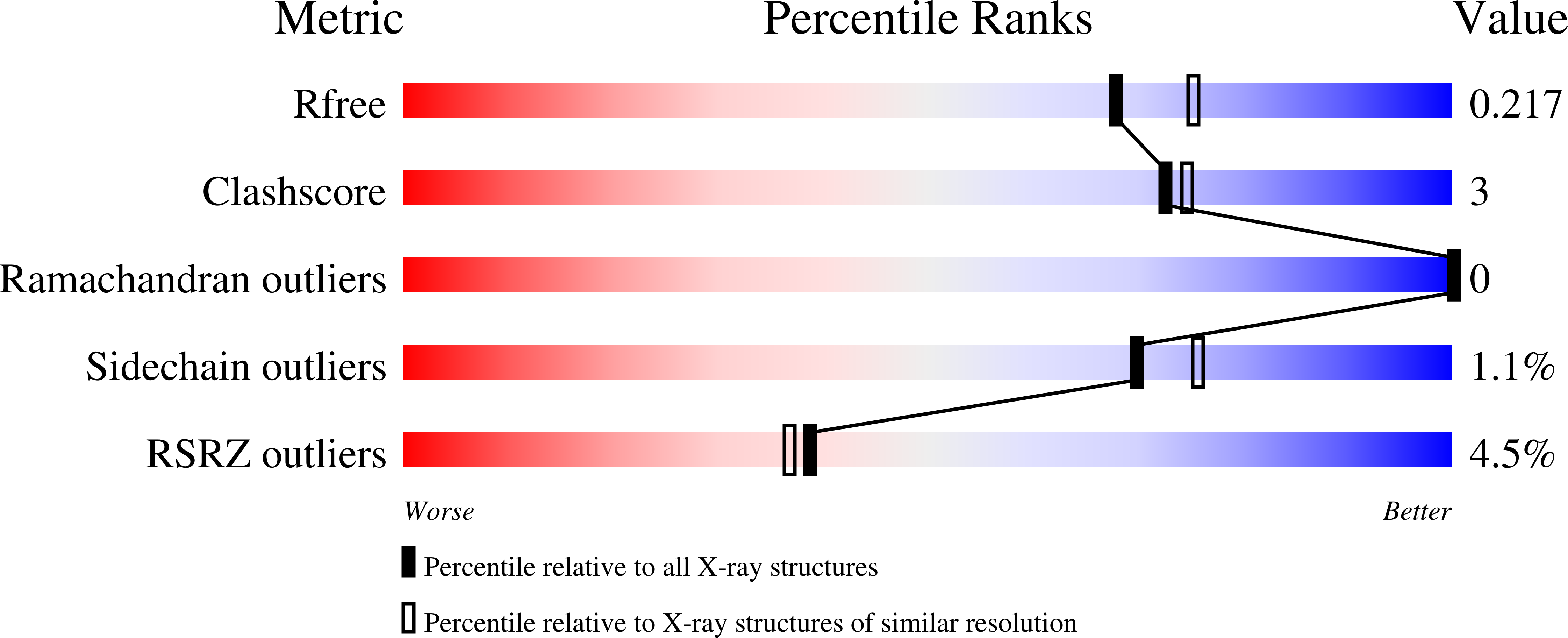
Resolution:
2.00 Å
R-Value Free:
0.21
R-Value Work:
0.18
R-Value Observed:
0.19
Space Group:
P 31 2 1
Macromolecular Entities
Polymer Type:polypeptide(L)
Description:NysL
Chain IDs:A, B
Chain Length:394
Number of Molecules:2
Biological Source:Streptomyces noursei
Primary Citation
Crystal structure of cytochrome P450 NysL and the structural basis for stereo- and regio-selective oxidation of antifungal macrolides.
J.Biol.Chem. 301 108185 108185 (2025)
PMID: 39814230 DOI: 10.1016/j.jbc.2025.108185

Abstact

NysL, a cytochrome P450 monooxygenase from the Gram-positive bacterium Streptomyces noursei, catalyzes the C10 hydroxylation of 10-deoxynystain to nystatin A1, a clinically important antifungal. In this study, we present the 2.0 Å resolution crystal structure of NysL bound to nystatin A1. The structure of this complex provides key insights into the structural elements that dictate the regio- and stereo-selective oxidation of large 20-44-membered macrolide substrates. The closely related AmphL operates on a similar 38-member macrolide but oxidizes C8 rather than C10. This difference requires that the substrate for AmphL penetrate further into the active site relative to NysL. The depth of substrate penetration is controlled by interactions between an area of the substrate binding pocket deemed the "back-wall" and the hemiketal ring of the macrolide substrate.

Legend

Protein

Chemical

Disease

Primary Citation of related structures