Deposition Date
2024-07-17
Release Date
2024-12-25
Last Version Date
2025-03-05
Entry Detail
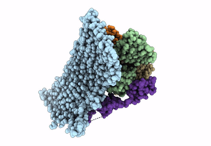
Biological Source:
Source Organism(s):
Escherichia coli (Taxon ID: 562)
Mus musculus (Taxon ID: 10090)
Camelus bactrianus (Taxon ID: 9837)
Homo sapiens (Taxon ID: 9606)
Mus musculus (Taxon ID: 10090)
Camelus bactrianus (Taxon ID: 9837)
Homo sapiens (Taxon ID: 9606)
Expression System(s):
Method Details:
Experimental Method:
Resolution:
3.73 Å
Aggregation State:
PARTICLE
Reconstruction Method:
SINGLE PARTICLE