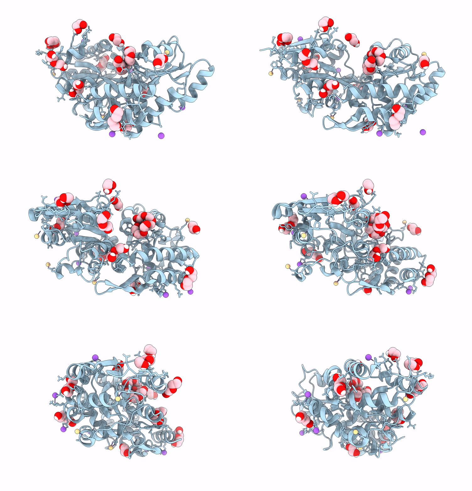
9CLD image
Entry Detail
PDB ID:
9CLD
Title:
Crystal structure of maltose binding protein (Apo)
Biological Source:
Source Organism:
Host Organism:
PDB Version:
Deposition Date:
2024-07-10
Release Date:
2024-12-18
Method Details:
Experimental Method:
Resolution:
1.58 Å
R-Value Free:
0.17
R-Value Work:
0.15
R-Value Observed:
0.15
Space Group:
P 1 21 1
Macromolecular Entities
Polymer Type:polypeptide(L)
Description:Maltose/maltodextrin-binding periplasmic protein
Mutations:T356A
Chain IDs:A
Chain Length:373
Number of Molecules:1
Biological Source:Escherichia coli
Primary Citation
Rendering Proteins Fluorescent Inconspicuously: Genetically Encoded 4-Cyanotryptophan Conserves Their Structure and Enables the Detection of Ligand Binding Sites.
Angew.Chem.Int.Ed.Engl. 64 e202421000 e202421000 (2025)
PMID: 39632265 DOI: 10.1002/anie.202421000

Abstact

Cyanotryptophans (CN-Trp) are privileged multimodal reporters on protein structure. They are similar in size to the canonical amino acid tryptophan and some of them exhibit bright fluorescence which responds sensitively to changes in the environment. We selected aminoacyl-tRNA synthetases specific for 4-, 5-, 6-, and 7-CN-Trp for high-yield in vivo production of proteins with a single, site-specifically introduced nitrile label. The absorption maximum of 4-CN-Trp is distinct from Trp, allowing the selective excitation of its intense fluorescence. 4-CN-Trp fluoresces in the visible range with an intensity rivalling that of 7-hydroxy-coumarin. Crystal structures of maltose binding protein demonstrate near-complete structural conservation when a native buried Trp residue is replaced by 4-CN-Trp. Besides presenting an inconspicuous tag for live cell microscopy, the intense fluorescence of 4-CN-Trp enables measurements of subnanomolar ligand binding affinities in isotropic solution, as demonstrated by the complex between rapamycin and the peptidyl-prolyl isomerase FKBP12 furnished with a 4-CN-Trp residue in the substrate binding pocket. Furthermore, 4-CN-Trp residues positioned at different locations of a protein containing multiple tryptophan residues permits using fluorescence quenching experiments to detect the proximity of individual Trp residues to the binding site of aromatic ligands.

Legend

Protein

Chemical

Disease

Primary Citation of related structures