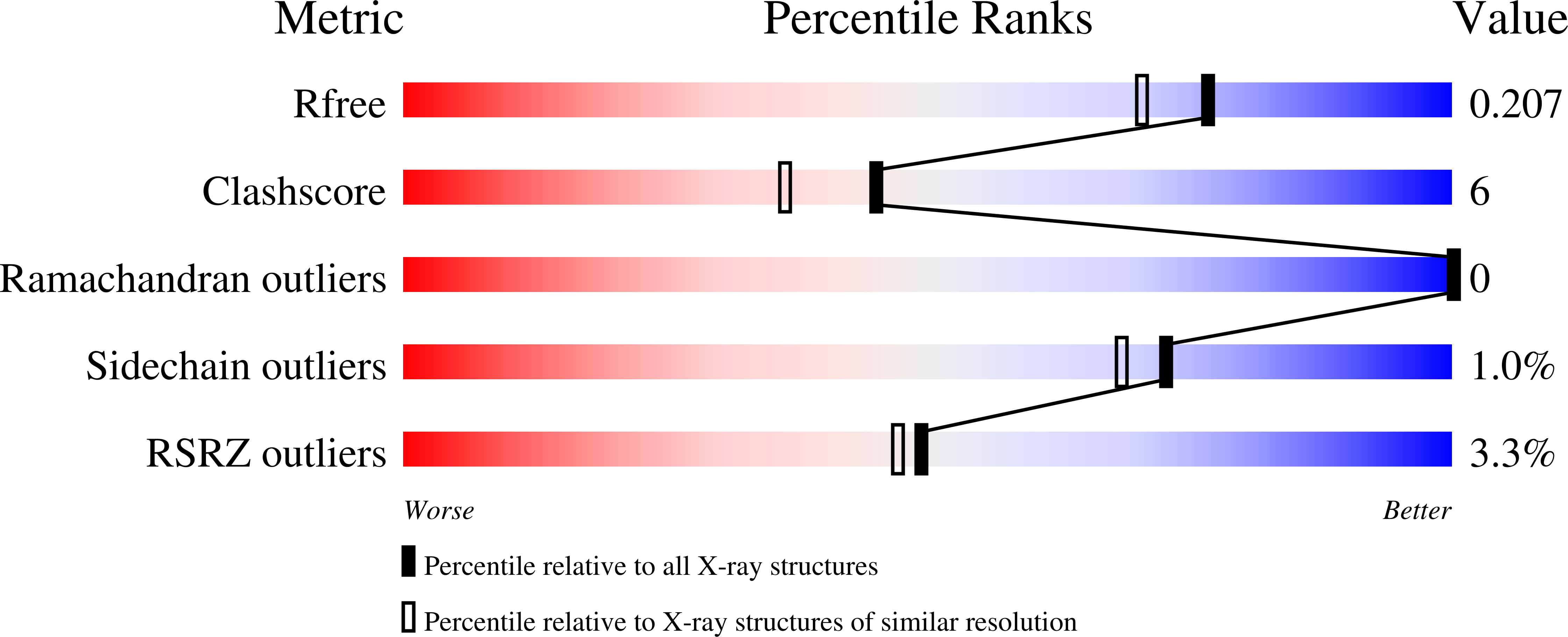
9CJ4 image
Deposition Date 2024-07-05
Release Date 2024-11-13
Last Version Date 2024-11-13
Entry Detail
PDB ID:
9CJ4
Keywords:
Title:
Dual phosphorylated human p38 alpha bound to BIRB796
Biological Source:
Source Organism:
Homo sapiens (Taxon ID: 9606)
Host Organism:
Method Details:
Experimental Method:
Resolution:
1.80 Å
R-Value Free:
0.19
R-Value Work:
0.15
R-Value Observed:
0.16
Space Group:
C 1 2 1
Macromolecular Entities
Polymer Type:polypeptide(L)
Molecule:Mitogen-activated protein kinase 14
Gene (Uniprot):MAPK14
Chain IDs:A
Chain Length:360
Number of Molecules:1
Biological Source:Homo sapiens
Primary Citation
Dual-Action Kinase Inhibitors Influence p38 alpha MAP Kinase Dephosphorylation.
Biorxiv ? ? ? (2024)
PMID: 39149408 DOI: 10.1101/2024.05.15.594272

Abstact

Reversible protein phosphorylation directs essential cellular processes including cell division, cell growth, cell death, inflammation, and differentiation. Because protein phosphorylation drives diverse diseases, kinases and phosphatases have been targets for drug discovery, with some achieving remarkable clinical success. Most protein kinases are activated by phosphorylation of their activation loops, which shifts the conformational equilibrium of the kinase towards the active state. To turn off the kinase, protein phosphatases dephosphorylate these sites, but how the conformation of the dynamic activation loop contributes to dephosphorylation was not known. To answer this, we modulated the activation loop conformational equilibrium of human p38α ΜΑP kinase with existing kinase inhibitors that bind and stabilize specific inactive activation loop conformations. From this, we discovered three inhibitors that increase the rate of dephosphorylation of the activation loop phospho-threonine by the PPM serine/threonine phosphatase WIP1. Hence, these compounds are "dual-action" inhibitors that simultaneously block the active site and stimulate p38α dephosphorylation. Our X-ray crystal structures of phosphorylated p38α bound to the dual-action inhibitors reveal a shared flipped conformation of the activation loop with a fully accessible phospho-threonine. In contrast, our X-ray crystal structure of phosphorylated apo human p38α reveals a different activation loop conformation with an inaccessible phospho-threonine, thereby explaining the increased rate of dephosphorylation upon inhibitor binding. These findings reveal a conformational preference of phosphatases for their targets and suggest a new approach to achieving improved potency and specificity for therapeutic kinase inhibitors.

Legend

Protein

Chemical

Disease

Primary Citation of related structures