Deposition Date
2024-06-27
Release Date
2024-09-11
Last Version Date
2024-10-02
Entry Detail
PDB ID:
9CF3
Keywords:
Title:
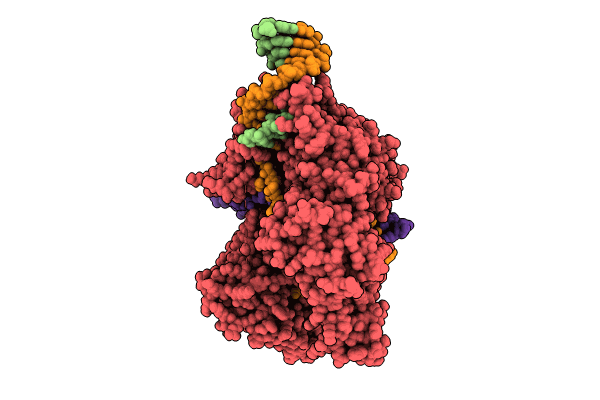
Parasitella parasitica Fanzor (PpFz) State 4
Biological Source:
Source Organism(s):
Parasitella parasitica (Taxon ID: 35722)
Saccharomyces cerevisiae (Taxon ID: 4932)
synthetic construct (Taxon ID: 32630)
Saccharomyces cerevisiae (Taxon ID: 4932)
synthetic construct (Taxon ID: 32630)
Expression System(s):
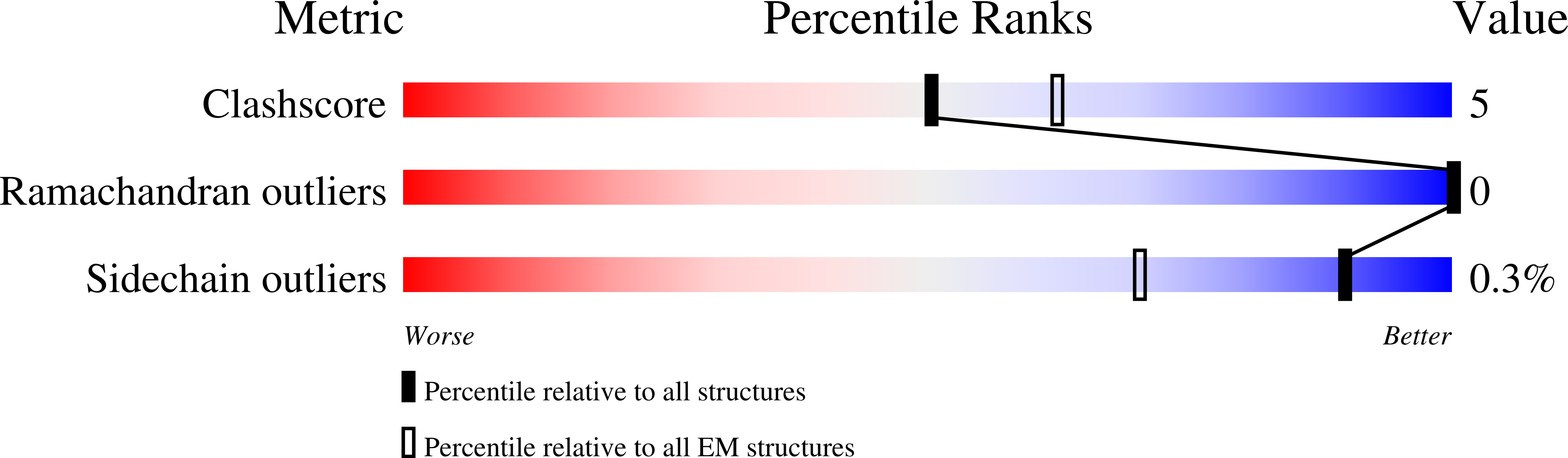
Method Details:
Experimental Method:
Resolution:
3.20 Å
Aggregation State:
PARTICLE
Reconstruction Method:
SINGLE PARTICLE