9CA3 image
Entry Detail
PDB ID:
9CA3
Keywords:
Title:
Crystal structure of MarE C280S in complex with cyanide bound heme and its native substrate, beta-methyl-L-tryptophan
Biological Source:
Source Organism:
PDB Version:
Deposition Date:
2024-06-16
Release Date:
2025-04-02
Method Details:
Experimental Method:
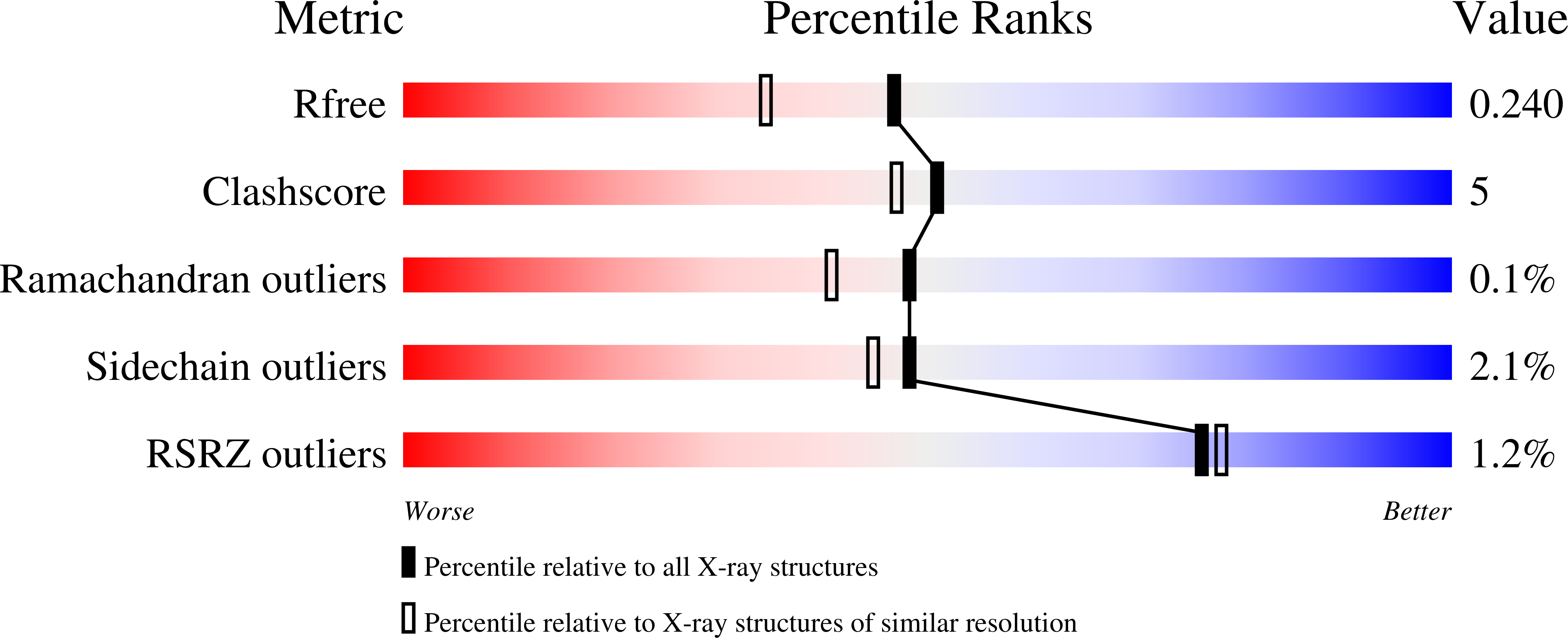
Resolution:
1.89 Å
R-Value Free:
0.24
R-Value Work:
0.19
R-Value Observed:
0.19
Space Group:
P 1 21 1
Macromolecular Entities
Polymer Type:polypeptide(L)
Description:Tryptophan 2,3-dioxygenase
Mutations:C280S
Chain IDs:A, B, C, D
Chain Length:286
Number of Molecules:4
Biological Source:Streptomyces sp. B9173
Primary Citation
Structural insights into 2-oxindole-forming monooxygenase MarE: Divergent architecture and substrate positioning versus tryptophan dioxygenases.
J.Biol.Chem. 301 108241 108241 (2025)
PMID: 39880093 DOI: 10.1016/j.jbc.2025.108241

Abstact

MarE, a heme-dependent enzyme, catalyzes a unique 2-oxindole-forming monooxygenation reaction from tryptophan metabolites. To elucidate its enzyme-substrate interaction mode, we present the first X-ray crystal structures of MarE in complex with its prime substrate, (2S,3S)-β-methyl-l-tryptophan and cyanide at 1.89 Å resolution as well as a truncated yet catalytically active version in complex with the substrate at 2.45 Å resolution. These structures establish MarE as a member of the heme-dependent aromatic oxygenase (HDAO) superfamily and reveal its evolutionary link to indoleamine 2,3-dioxygenase (IDO) and tryptophan 2,3-dioxygenase (TDO). While MarE adopts a global structure resembling the homotetrameric TDO, it features a simplified α6 helix compared to TDO's more elaborate αE and αH helices with additional αF and αG regions. Despite differing oxygen activation outcomes, MarE shares a substrate binding mode similar to IDO and TDO, with the indole nitrogen of its substrate oriented toward the heme iron in the ternary cyano complex, interacting with His55. The substrate's carboxylate group engages Arg118, with mutational studies confirming the roles of these residues in substrate binding. However, the second-sphere interactions with the substrate's α-amino nitrogen differ between MarE and TDO, and the substrate's orientation in the binary complex remains ambiguous due to two possible conformations. Notably, TDO features an extensive hydrogen-bonding network around the heme propionate below the heme plane, which is absent in MarE, suggesting mechanistic differences. These structural insights lay a foundation for further mechanistic studies, particularly for understanding how heme-dependent enzymes oxygenate tryptophan-derived metabolites.

Legend

Protein

Chemical

Disease

Primary Citation of related structures