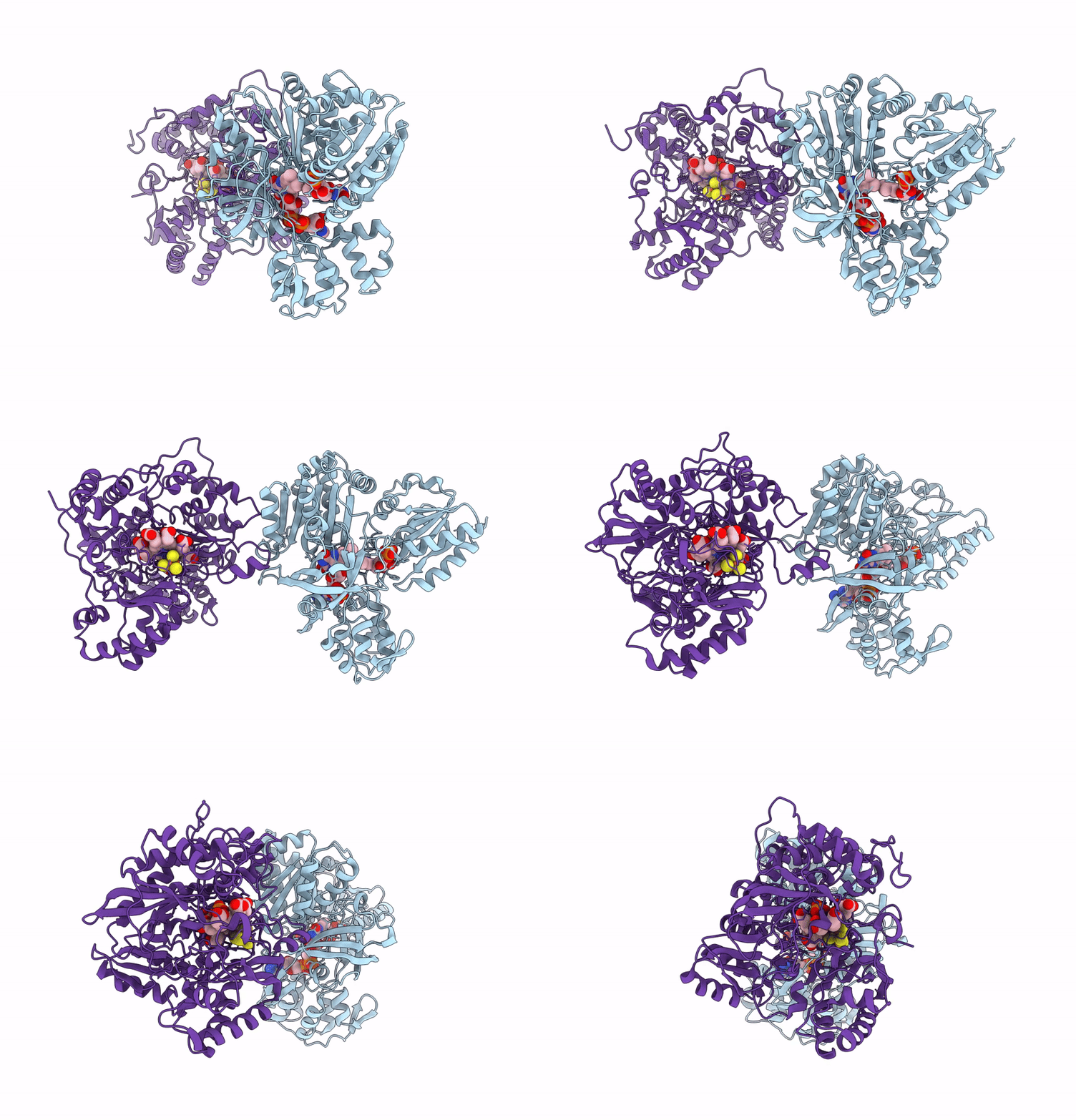
9C91 image
Deposition Date 2024-06-13
Release Date 2025-02-12
Last Version Date 2025-08-27
Entry Detail
PDB ID:
9C91
Keywords:
Title:
Assimilatory NADPH-dependent sulfite reductase minimal dimer
Biological Source:
Source Organism:
Escherichia coli (Taxon ID: 562)
Host Organism:
Method Details:
Experimental Method:
Resolution:
2.78 Å
Aggregation State:
PARTICLE
Reconstruction Method:
SINGLE PARTICLE
Macromolecular Entities
Structures with similar UniProt ID
Protein Blast
Polymer Type:polypeptide(L)
Molecule:Sulfite reductase [NADPH] flavoprotein alpha-component
Chain IDs:A
Chain Length:575
Number of Molecules:1
Biological Source:Escherichia coli
Structures with similar UniProt ID
Protein Blast
Polymer Type:polypeptide(L)
Molecule:Sulfite reductase [NADPH] hemoprotein beta-component
Chain IDs:B
Chain Length:570
Number of Molecules:1
Biological Source:Escherichia coli
Primary Citation
Structure of dimerized assimilatory NADPH-dependent sulfite reductase reveals the minimal interface for diflavin reductase binding.
Nat Commun 16 2955 2955 (2025)
PMID: 40140349 DOI: 10.1038/s41467-025-58037-5

Abstact

Escherichia coli NADPH-dependent assimilatory sulfite reductase (SiR) reduces sulfite by six electrons to make sulfide for incorporation into sulfur-containing biomolecules. SiR has two subunits: an NADPH, FMN, and FAD-binding diflavin flavoprotein and a siroheme/Fe4S4 cluster-containing hemoprotein. The molecular interactions that govern subunit binding have been unknown since the discovery of SiR over 50 years ago because SiR is flexible, thus has been intransigent for traditional high-resolution structural analysis. We use a combination of the chameleon® plunging system with a fluorinated lipid to overcome the challenges of preserving a flexible molecule to determine a 2.78 Å-resolution cryo-EM structure of a minimal heterodimer complex. Chameleon®, combined with the fluorinated lipid, overcomes persistent denaturation at the air-water interface. Using a previously characterized minimal heterodimer reduces the heterogeneity of a structurally heterogeneous complex to a level that we analyze using multi-conformer cryo-EM image analysis algorithms. Here, we report the near-atomic resolution structure of the flavoprotein/hemoprotein complex, revealing how they interact in a minimal interface. Further, we determine the structural elements that discriminate between pairing a hemoprotein with a diflavin reductase, as in the E. coli homolog, or a ferredoxin partner, as in maize (Zea mays).

Legend

Protein

Chemical

Disease

Primary Citation of related structures