9C66 image
Deposition Date 2024-06-07
Release Date 2024-08-28
Last Version Date 2024-10-23
Entry Detail
PDB ID:
9C66
Title:
Structure of the Mena EVH1 domain bound to the polyproline segment of PTP1B
Biological Source:
Source Organism:
Homo sapiens (Taxon ID: 9606)
Method Details:
Experimental Method:
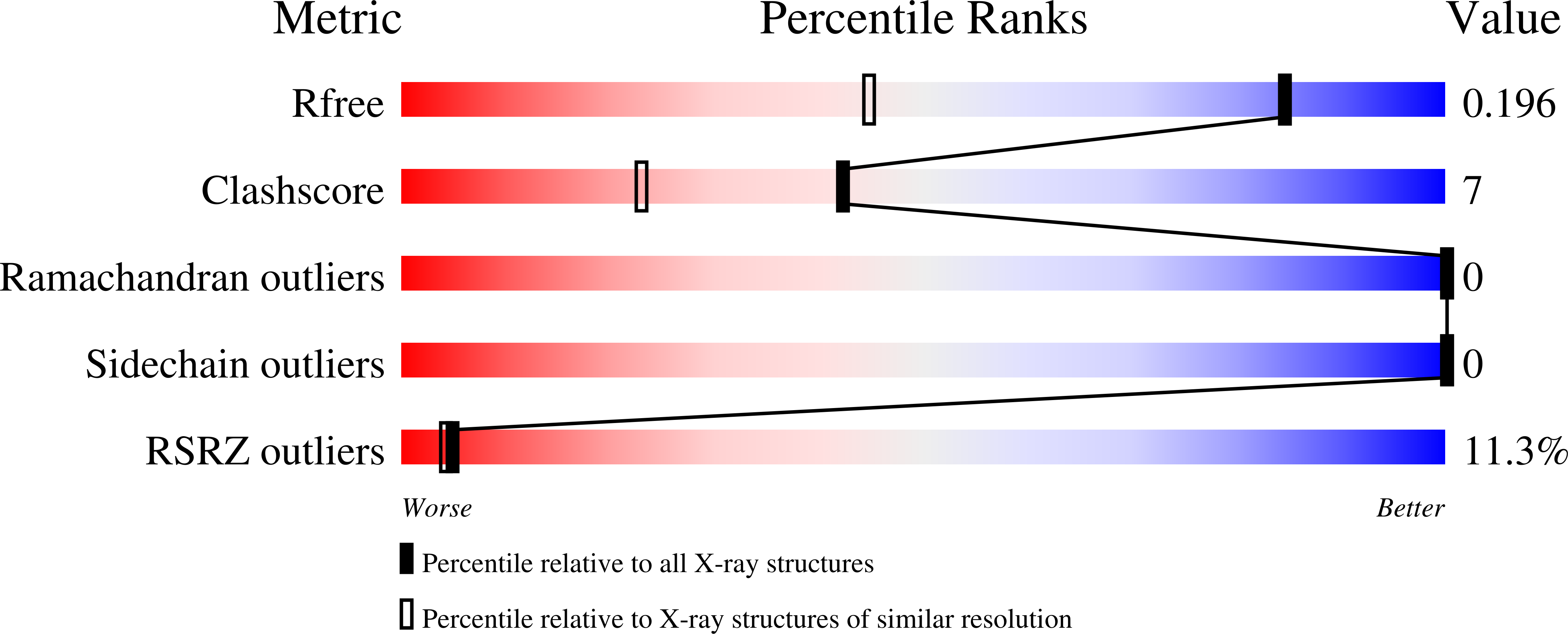
Resolution:
1.40 Å
R-Value Free:
0.19
R-Value Work:
0.18
R-Value Observed:
0.18
Space Group:
P 21 21 21
Macromolecular Entities
Structures with similar UniProt ID
Protein Blast
Polymer Type:polypeptide(L)
Molecule:Protein enabled homolog
Gene (Uniprot):ENAH
Chain IDs:A
Chain Length:114
Number of Molecules:1
Biological Source:Homo sapiens
Structures with similar UniProt ID
Protein Blast
Polymer Type:polypeptide(L)
Molecule:poly-proline segment of PTP1B
Gene (Uniprot):PTPN1
Chain IDs:B
Chain Length:10
Number of Molecules:1
Biological Source:Homo sapiens
Modified Residue
Compound ID Chain ID Parent Comp ID Details 2D Image
CME A CYS modified residue
CSO A CYS modified residue
Primary Citation
Insights into the Interaction Landscape of the EVH1 Domain of Mena.
Biochemistry 63 2183 2195 (2024)
PMID: 39138154 DOI: 10.1021/acs.biochem.4c00331

Abstact

The Enabled/VASP homology 1 (EVH1) domain is a small module that interacts with proline-rich stretches in its ligands and is found in various signaling and scaffolding proteins. Mena, the mammalian homologue of Ena, is involved in diverse actin-associated events, such as membrane dynamics, bacterial motility, and tumor intravasation and extravasation. Two-dimensional (2D) 1H-15N HSQC NMR was used to study Mena EVH1 binding properties, defining the amino acids involved in ligand recognition for the physiological ligands ActA and PCARE, and a synthetic polyproline-inspired small molecule (hereafter inhibitor 6c). Chemical shift perturbations indicated that proline-rich segments bind in the conserved EVH1 hydrophobic cleft. The PCARE-derived peptide elicited more perturbations compared to the ActA-derived peptide, consistent with a previous report of a structural alteration in the solvent-exposed β7-β8 loop. Unexpectedly, EVH1 and the proline-rich segment of PTP1B did not exhibit NMR chemical shift perturbations; however, the high-resolution crystal structure implicated the conserved EVH1 hydrophobic cleft in ligand recognition. Intrinsic steady-state fluorescence and fluorescence polarization assays indicate that residues outside the proline-rich segment enhance the ligand affinity for EVH1 (Kd = 3-8 μM). Inhibitor 6c displayed tighter binding (Kd ∼ 0.3 μM) and occupies the same EVH1 cleft as physiological ligands. These studies revealed that the EVH1 domain enhances ligand affinity through recognition of residues flanking the proline-rich segments. Additionally, a synthetic inhibitor binds more tightly to the EVH1 domain than natural ligands, occupying the same hydrophobic cleft.

Legend

Protein

Chemical

Disease

Primary Citation of related structures
Feedback Form
Name
Email
Institute
Feedback