Deposition Date
2024-06-06
Release Date
2024-06-26
Last Version Date
2024-10-16
Entry Detail
PDB ID:
9C5S
Keywords:
Title:
Disulfide-linked, antiparallel p53-derived peptide dimer (CV1)
Biological Source:
Source Organism(s):
Homo sapiens (Taxon ID: 9606)
Method Details:
Experimental Method:
Resolution:
1.01 Å
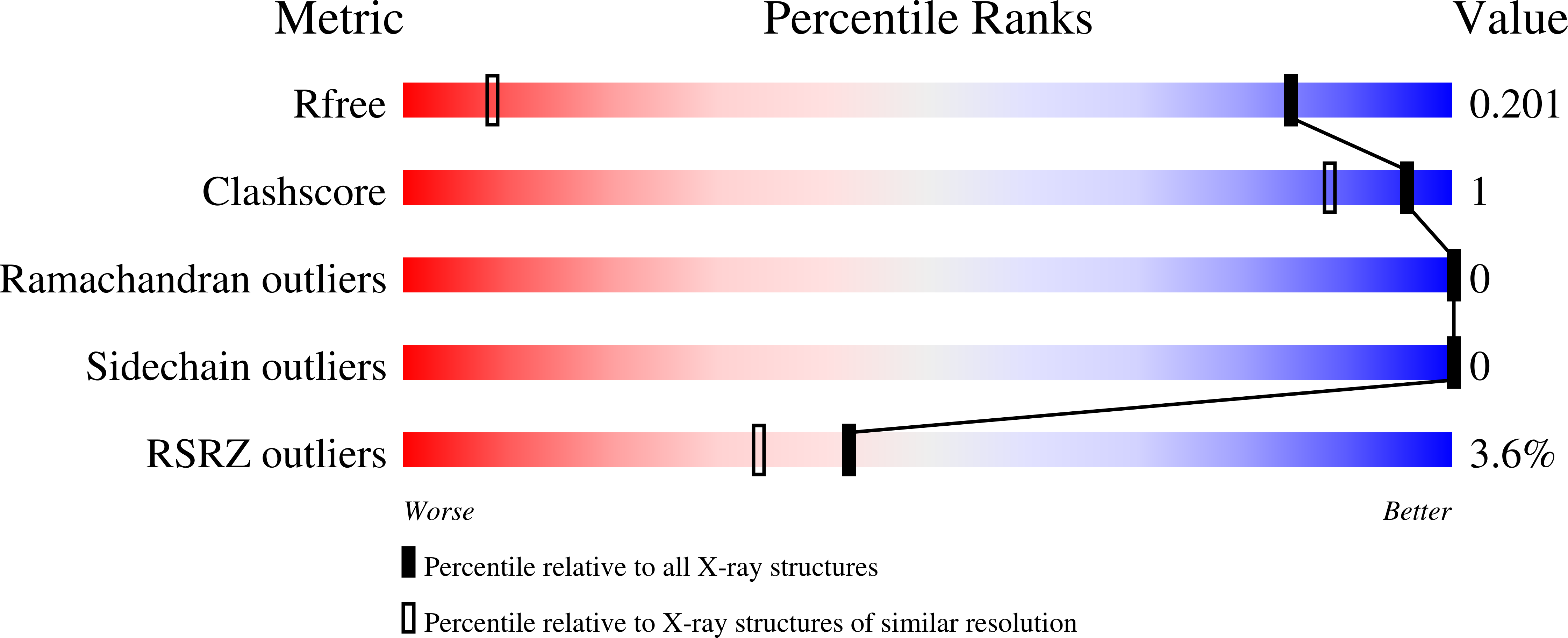
R-Value Free:
0.20
R-Value Work:
0.19
R-Value Observed:
0.19
Space Group:
P 31 2 1