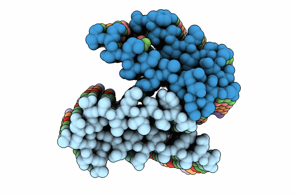
9C1U image
Entry Detail
PDB ID:
9C1U
EMDB ID:
Keywords:
Title:
Cryo-EM Structure of a Tm1C Fibril
Biological Source:
Source Organism:
PDB Version:
Deposition Date:
2024-05-29
Release Date:
2024-10-09
Method Details:
Experimental Method:
Resolution:
2.31 Å
Aggregation State:
HELICAL ARRAY
Reconstruction Method:
HELICAL
Macromolecular Entities
Polymer Type:polypeptide(L)
Description:Tropomyosin 1 I/C
Chain IDs:A, B, C, D, E, F, G, H, I, J, K, L
Chain Length:71
Number of Molecules:12
Biological Source:Drosophila melanogaster
Ligand Molecules
Primary Citation
Cryo-EM and solid state NMR together provide a more comprehensive structural investigation of protein fibrils.
Protein Sci. 33 e5168 e5168 (2024)
PMID: 39276003 DOI: 10.1002/pro.5168

Abstact

The tropomyosin 1 isoform I/C C-terminal domain (Tm1-LC) fibril structure is studied jointly with cryogenic electron microscopy (cryo-EM) and solid state nuclear magnetic resonance (NMR). This study demonstrates the complementary nature of these two structural biology techniques. Chemical shift assignments from solid state NMR are used to determine the secondary structure at the level of individual amino acids, which is faithfully seen in cryo-EM reconstructions. Additionally, solid state NMR demonstrates that the region not observed in the reconstructed cryo-EM density is primarily in a highly mobile random coil conformation rather than adopting multiple rigid conformations. Overall, this study illustrates the benefit of investigations combining cryo-EM and solid state NMR to investigate protein fibril structure.

Legend

Protein

Chemical

Disease

Primary Citation of related structures