
Deposition Date
2024-05-07
Release Date
2025-05-14
Last Version Date
2025-05-14
Entry Detail
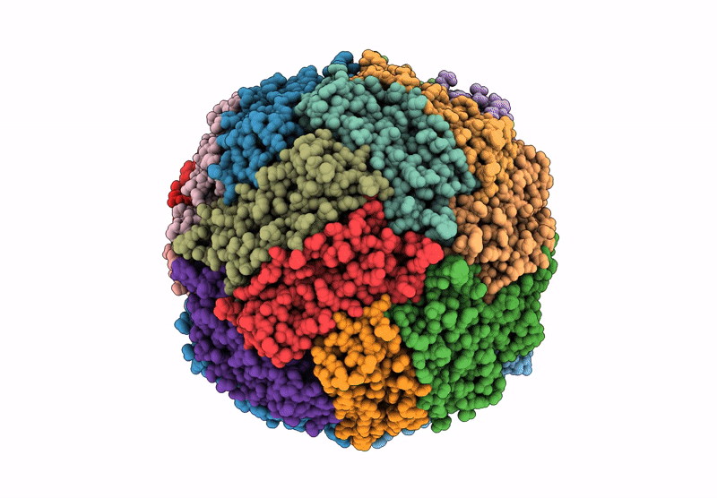
PDB ID:
9BPJ
Keywords:
Title:
Human light chain ferritin reacted with iron (3 Fe2+ to ferritin monomer ratio). Reconstruction of particles with one nanoparticle
Biological Source:
Source Organism(s):
Homo sapiens (Taxon ID: 9606)
Expression System(s):
Method Details:
Experimental Method:
Resolution:
2.85 Å
Aggregation State:
PARTICLE
Reconstruction Method:
SINGLE PARTICLE
