
Deposition Date
2024-05-07
Release Date
2025-03-26
Last Version Date
2025-08-13
Entry Detail
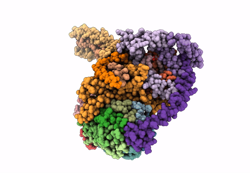
PDB ID:
9BPG
Keywords:
Title:
Artemia franciscana ATP synthase FO domain, state 1, pH 7.0
Biological Source:
Source Organism(s):
Artemia franciscana (Taxon ID: 6661)
Method Details:
Experimental Method:
Resolution:
3.30 Å
Aggregation State:
PARTICLE
Reconstruction Method:
SINGLE PARTICLE
