Deposition Date
2024-04-30
Release Date
2025-04-30
Last Version Date
2025-04-30
Entry Detail
PDB ID:
9BLJ
Keywords:
Title:
Crystal structure of a serine protease inhibitor HPI from Hevea brasiliensis
Biological Source:
Source Organism(s):
Hevea brasiliensis (Taxon ID: 3981)
Expression System(s):
Method Details:
Experimental Method:
Resolution:
1.74 Å
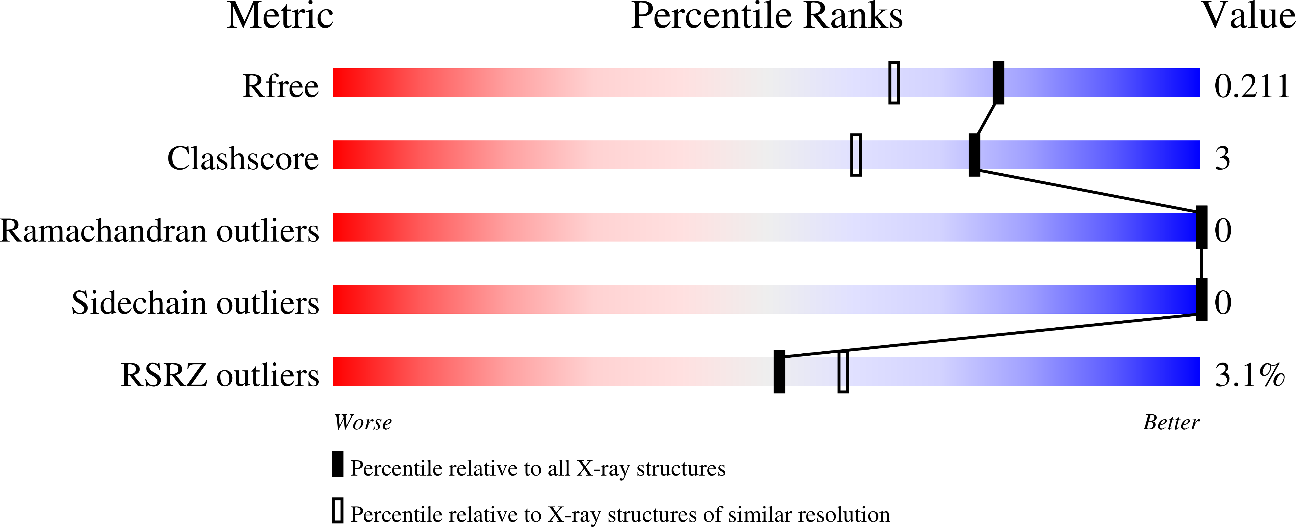
R-Value Free:
0.21
R-Value Work:
0.19
R-Value Observed:
0.19
Space Group:
P 61 2 2