9BKW image
Deposition Date 2024-04-29
Release Date 2024-05-08
Last Version Date 2025-12-10
Entry Detail
PDB ID:
9BKW
Title:
Crystal structure of a C2 domain from Trichomonas vaginalis
Biological Source:
Source Organism:
Method Details:
Experimental Method:
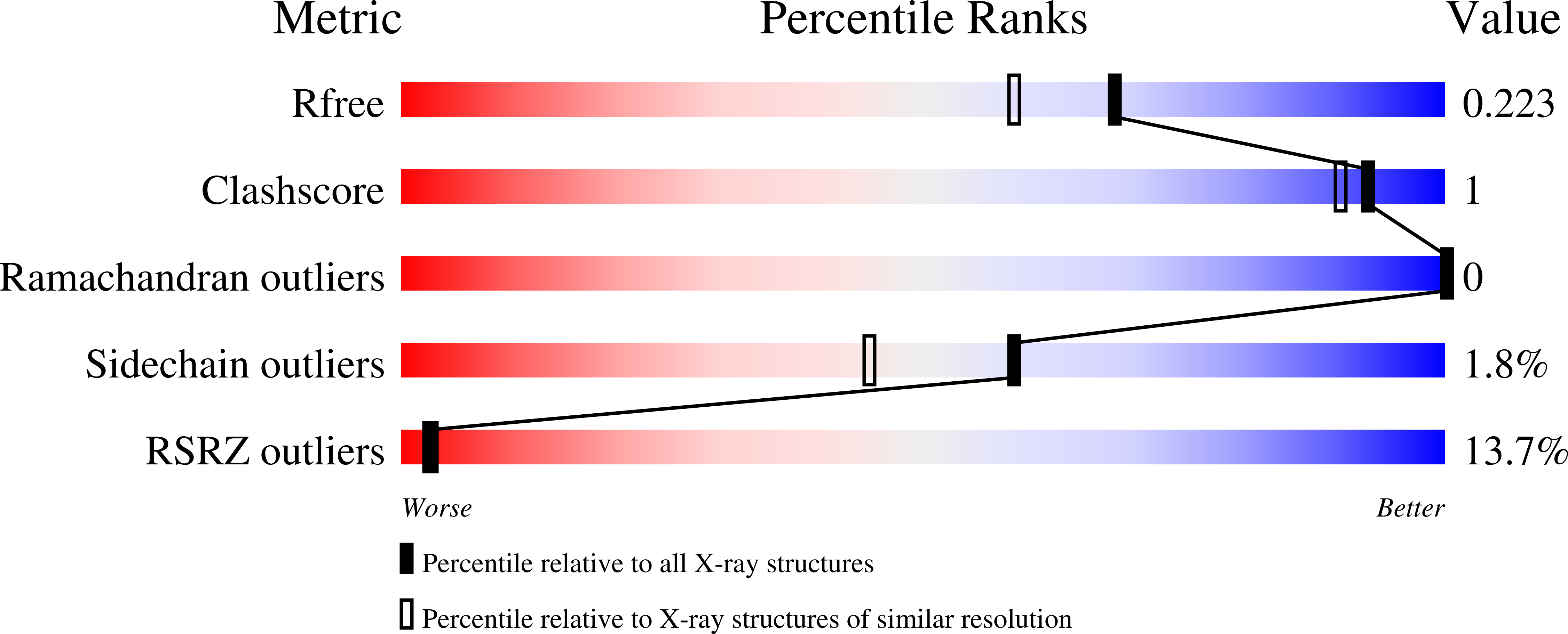
Resolution:
1.85 Å
R-Value Free:
0.22
R-Value Work:
0.19
R-Value Observed:
0.19
Space Group:
P 41 21 2
Macromolecular Entities
Structures with similar UniProt ID
Protein Blast
Polymer Type:polypeptide(L)
Molecule:XYPPX repeat family protein
Gene (Uniprot):TVAG_347440
Chain IDs:A
Chain Length:138
Number of Molecules:1
Biological Source:Trichomonas vaginalis G3
Primary Citation
A Trichomonas vaginalis C2-XYPPX-repeat protein with a structured C2 domain displaying dampened flexibility upon binding calcium.
Biochimie ? ? ? (2025)
PMID: 41285221 DOI: 10.1016/j.biochi.2025.11.011

Abstact

C2 domains are ubiquitous membrane-binding modules of ∼130 residues in eukaryotes that are often associated with proteins involved in membrane trafficking and lipid modification. The genome of Trichomonas vaginalis, the most common, non-viral, sexually transmitted human pathogen, encodes eight genes that contain a N-terminal C2 module linked to a XYPPX-repeat domain of more than four XYPPX repeats (C2-XYPPX). While the function of the XYPPX-repeat domain remains unknown, its multiple association with C2 domains in T. vaginalis suggests it is important. The C2 domain from one of these C2-XYPPX-repeat proteins, Tv-C2-1, was structurally and physically characterized using X-ray crystallography and NMR spectroscopy. The crystal structure for Tv-C2-1 shows that this domain shares a fold common to all C2 domains, a compact Greek-key motif composed of eight anti-parallel β-strands in the type-2 topology. An NMR chemical shift perturbation study with Ca2+ showed that Tv-C2-1 bound two Ca2+ atoms primarily via two loops (loop-1 and loop-3) on the predicted calcium binding face of the protein with Kds of 58.0 ± 0.1 μM and 232 ± 6 μM. Estimations of the overall rotational correlation time, τc, in the apo (11.1 ns) and Ca2+-bound (9.2 ns) state suggests the protein becomes more compact upon Ca2+ binding, consistent with a decrease in dynamics in loop-3 and marginally in loop-1 suggested by amide 15N heteronuclear steady-state {1H}-15N NOEs. Showing Tv-C2-1 binds calcium and adopts a compact Greek-key motif structure, two primary features of C2 domains, suggests understanding the function of the XYPPX-repeat domain may be warranted.

Legend

Protein

Chemical

Disease

Primary Citation of related structures
Feedback Form
Name
Email
Institute
Feedback