Deposition Date
2024-04-24
Release Date
2024-07-17
Last Version Date
2024-11-13
Entry Detail
PDB ID:
9BIY
Keywords:
Title:
Crystal structure of the periplasmic domain of IgaA from Escherichia coli
Biological Source:
Source Organism:
Escherichia coli (Taxon ID: 562)
Host Organism:
Method Details:
Experimental Method:
Resolution:
1.80 Å
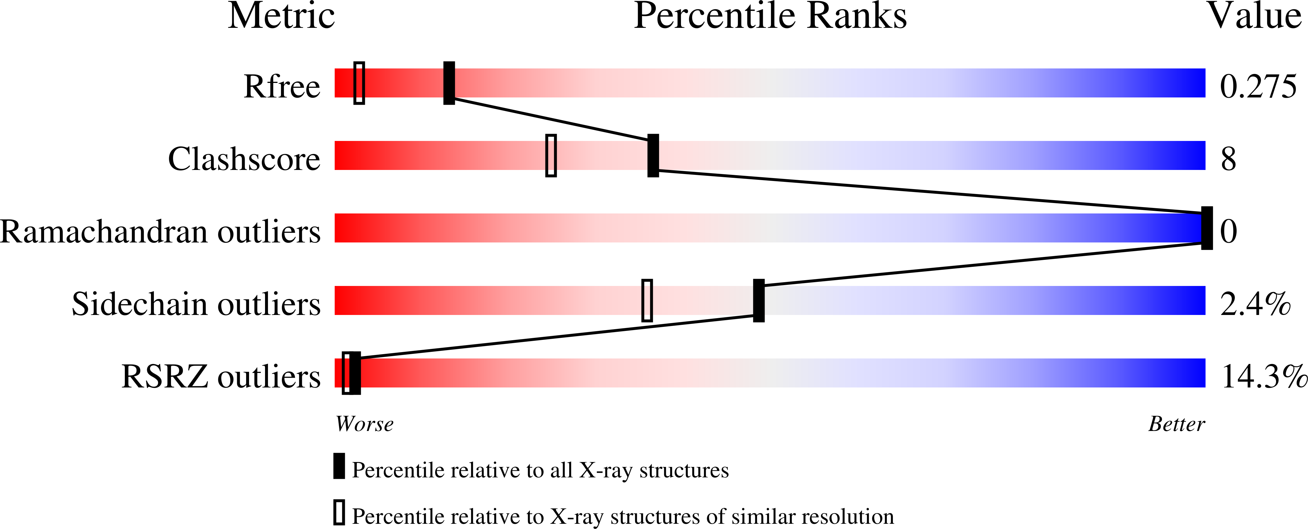
R-Value Free:
0.27
R-Value Work:
0.23
R-Value Observed:
0.24
Space Group:
C 1 2 1