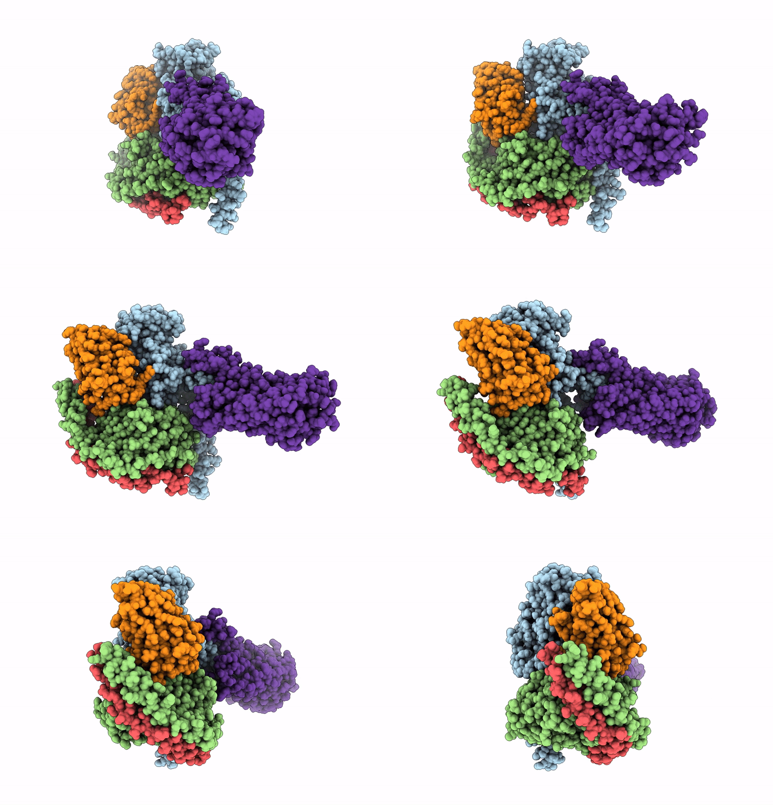
9BIP image
Entry Detail
PDB ID:
9BIP
EMDB ID:
Title:
Human proton sensing receptor GPR4 in complex with miniGs
Biological Source:
Source Organism:
Host Organism:
PDB Version:
Deposition Date:
2024-04-24
Release Date:
2025-01-15
Method Details:
Experimental Method:
Resolution:
2.80 Å
Aggregation State:
PARTICLE
Reconstruction Method:
SINGLE PARTICLE
Macromolecular Entities
Polymer Type:polypeptide(L)
Description:miniGs
Chain IDs:A
Chain Length:261
Number of Molecules:1
Biological Source:Homo sapiens
Polymer Type:polypeptide(L)
Description:Guanine nucleotide-binding protein G(I)/G(S)/G(T) subunit beta-1
Chain IDs:B
Chain Length:370
Number of Molecules:1
Biological Source:Homo sapiens
Polymer Type:polypeptide(L)
Description:Guanine nucleotide-binding protein G(I)/G(S)/G(O) subunit gamma-2
Chain IDs:C (auth: G)
Chain Length:68
Number of Molecules:1
Biological Source:Homo sapiens
Polymer Type:polypeptide(L)
Description:Nb35
Chain IDs:D (auth: N)
Chain Length:142
Number of Molecules:1
Biological Source:Lama glama
Polymer Type:polypeptide(L)
Description:G-protein coupled receptor 4
Chain IDs:E (auth: R)
Chain Length:373
Number of Molecules:1
Biological Source:Homo sapiens
Ligand Molecules
Primary Citation
Molecular basis of proton sensing by G protein-coupled receptors.
Cell 188 671 687.e20 (2025)
PMID: 39753132 DOI: 10.1016/j.cell.2024.11.036

Abstact

Three proton-sensing G protein-coupled receptors (GPCRs)-GPR4, GPR65, and GPR68-respond to extracellular pH to regulate diverse physiology. How protons activate these receptors is poorly understood. We determined cryogenic-electron microscopy (cryo-EM) structures of each receptor to understand the spatial arrangement of proton-sensing residues. Using deep mutational scanning (DMS), we determined the functional importance of every residue in GPR68 activation by generating ∼9,500 mutants and measuring their effects on signaling and surface expression. Constant-pH molecular dynamics simulations provided insights into the conformational landscape and protonation patterns of key residues. This unbiased approach revealed that, unlike other proton-sensitive channels and receptors, no single site is critical for proton recognition. Instead, a network of titratable residues extends from the extracellular surface to the transmembrane region, converging on canonical motifs to activate proton-sensing GPCRs. Our approach integrating structure, simulations, and unbiased functional interrogation provides a framework for understanding GPCR signaling complexity.

Legend

Protein

Chemical

Disease

Primary Citation of related structures