Deposition Date
2024-04-21
Release Date
2025-01-22
Last Version Date
2025-06-04
Entry Detail
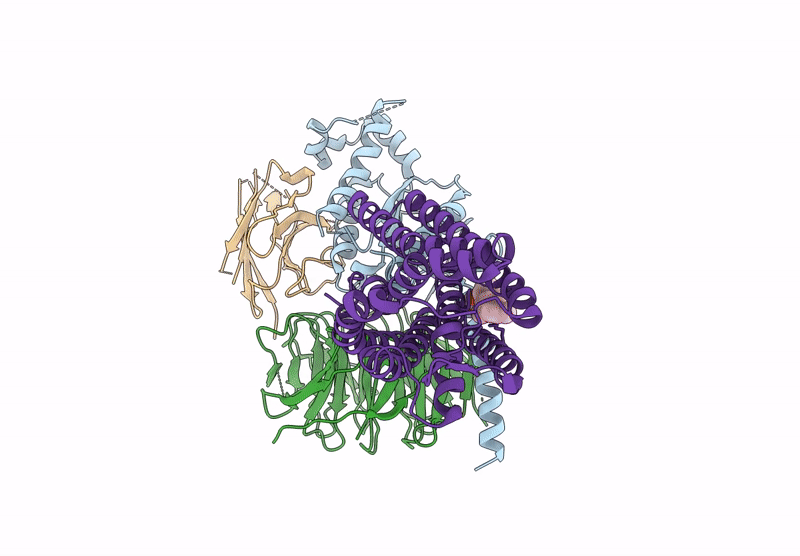
PDB ID:
9BHM
Keywords:
Title:
Human proton sensing receptor GPR68 in complex with miniGs
Biological Source:
Source Organism(s):
Homo sapiens (Taxon ID: 9606)
Lama glama (Taxon ID: 9844)
Lama glama (Taxon ID: 9844)
Expression System(s):
Method Details:
Experimental Method:
Resolution:
2.90 Å
Aggregation State:
PARTICLE
Reconstruction Method:
SINGLE PARTICLE