Deposition Date
2024-03-20
Release Date
2024-05-29
Last Version Date
2025-06-11
Entry Detail
PDB ID:
9B3R
EMDB ID:
Keywords:
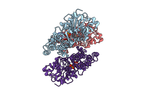
Title:
The structure of human cardiac F-actin
Biological Source:
Source Organism(s):
Homo sapiens (Taxon ID: 9606)
Expression System(s):
Method Details:
Experimental Method:
Resolution:
3.50 Å
Aggregation State:
FILAMENT
Reconstruction Method:
HELICAL