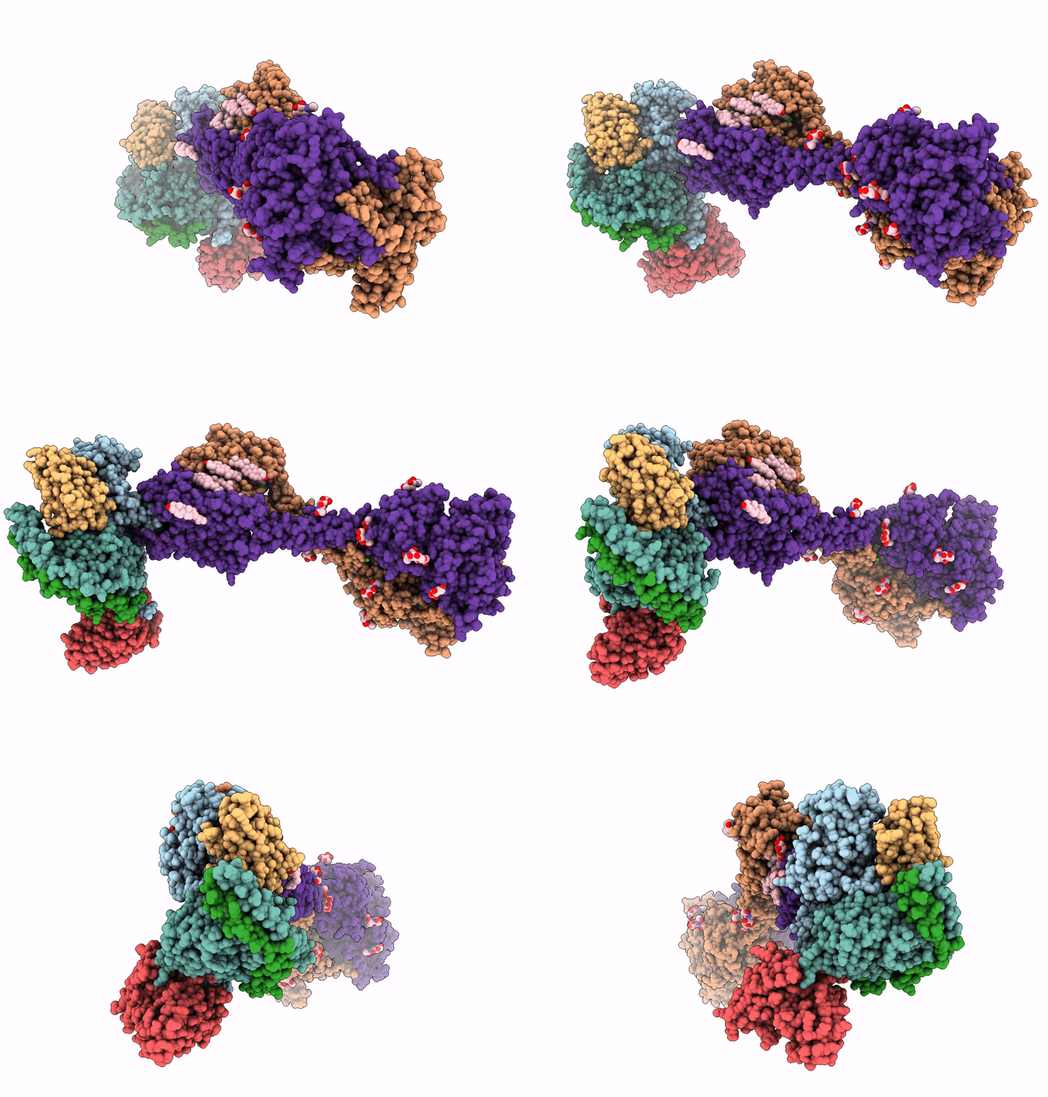
9AXF image
Entry Detail
PDB ID:
9AXF
EMDB ID:
Title:
Structure of human calcium-sensing receptor in complex with chimeric Gq (miniGisq) protein in detergent
Biological Source:
Host Organism:
PDB Version:
Deposition Date:
2024-03-06
Release Date:
2024-04-17
Method Details:
Experimental Method:
Resolution:
3.50 Å
Aggregation State:
PARTICLE
Reconstruction Method:
SINGLE PARTICLE
Macromolecular Entities
Polymer Type:polypeptide(L)
Description:Guanine nucleotide-binding protein G(i) subunit alpha-1,Adenylate cyclase-stimulating G alpha protein
Chain IDs:C (auth: A)
Chain Length:246
Number of Molecules:1
Biological Source:Homo sapiens
Polymer Type:polypeptide(L)
Description:Guanine nucleotide-binding protein G(I)/G(S)/G(T) subunit beta-1
Chain IDs:D (auth: B)
Chain Length:348
Number of Molecules:1
Biological Source:Homo sapiens
Polymer Type:polypeptide(L)
Description:Guanine nucleotide-binding protein G(I)/G(S)/G(O) subunit gamma-2
Chain IDs:E (auth: G)
Chain Length:71
Number of Molecules:1
Biological Source:Homo sapiens
Polymer Type:polypeptide(L)
Description:Single-chain antibody fragment scFv16
Chain IDs:F (auth: H)
Chain Length:297
Number of Molecules:1
Biological Source:Mus musculus
Polymer Type:polypeptide(L)
Description:Nanobody Nb-35
Chain IDs:G (auth: N)
Chain Length:160
Number of Molecules:1
Biological Source:Lama glama
Polymer Type:polypeptide(L)
Description:Extracellular calcium-sensing receptor
Chain IDs:A (auth: R), B (auth: Q)
Chain Length:911
Number of Molecules:2
Biological Source:Homo sapiens
Primary Citation
Promiscuous G-protein activation by the calcium-sensing receptor.
Nature 629 481 488 (2024)
PMID: 38632411 DOI: 10.1038/s41586-024-07331-1

Abstact

The human calcium-sensing receptor (CaSR) detects fluctuations in the extracellular Ca2+ concentration and maintains Ca2+ homeostasis1,2. It also mediates diverse cellular processes not associated with Ca2+ balance3-5. The functional pleiotropy of CaSR arises in part from its ability to signal through several G-protein subtypes6. We determined structures of CaSR in complex with G proteins from three different subfamilies: Gq, Gi and Gs. We found that the homodimeric CaSR of each complex couples to a single G protein through a common mode. This involves the C-terminal helix of each Gα subunit binding to a shallow pocket that is formed in one CaSR subunit by all three intracellular loops (ICL1-ICL3), an extended transmembrane helix 3 and an ordered C-terminal region. G-protein binding expands the transmembrane dimer interface, which is further stabilized by phospholipid. The restraint imposed by the receptor dimer, in combination with ICL2, enables G-protein activation by facilitating conformational transition of Gα. We identified a single Gα residue that determines Gq and Gs versus Gi selectivity. The length and flexibility of ICL2 allows CaSR to bind all three Gα subtypes, thereby conferring capacity for promiscuous G-protein coupling.

Legend

Protein

Chemical

Disease

Primary Citation of related structures