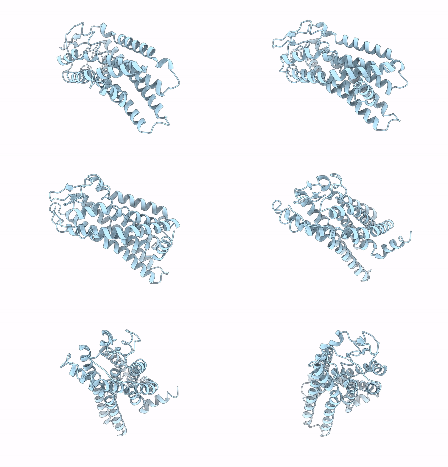
8ZFE image
Entry Detail
PDB ID:
8ZFE
EMDB ID:
Title:
Cryo-EM structure of the mmGPR4-Gs receptor in pH7.2
Biological Source:
Source Organism:
Host Organism:
PDB Version:
Deposition Date:
2024-05-07
Release Date:
2025-02-26
Method Details:
Experimental Method:
Resolution:
2.56 Å
Aggregation State:
PARTICLE
Reconstruction Method:
SINGLE PARTICLE
Macromolecular Entities
Polymer Type:polypeptide(L)
Description:G-protein coupled receptor 4
Chain IDs:A (auth: R)
Chain Length:365
Number of Molecules:1
Biological Source:Mus musculus
Ligand Molecules
Primary Citation
Evolutionary study and structural basis of proton sensing by Mus GPR4 and Xenopus GPR4.
Cell 188 653 670.e24 (2025)
PMID: 39753131 DOI: 10.1016/j.cell.2024.12.001

Abstact

Animals have evolved pH-sensing membrane receptors, such as G-protein-coupled receptor 4 (GPR4), to monitor pH changes related to their physiology and generate adaptive reactions. However, the evolutionary trajectory and structural mechanism of proton sensing by GPR4 remain unresolved. Here, we observed a positive correlation between the optimal pH of GPR4 activity and the blood pH range across different species. By solving 7-cryoelectron microscopy (cryo-EM) structures of Xenopus tropicalis GPR4 (xtGPR4) and Mus musculus GPR4 (mmGPR4) under varying pH conditions, we identified that protonation of HECL2-45.47 and H7.36 enabled polar network establishment and tighter association between the extracellular loop 2 (ECL2) and 7 transmembrane (7TM) domain, as well as a conserved propagating path, which are common mechanisms underlying protonation-induced GPR4 activation across different species. Moreover, protonation of distinct extracellular HECL2-45.41 contributed to the more acidic optimal pH range of xtGPR4. Overall, our study revealed common and distinct mechanisms of proton sensing by GPR4, from a structural, functional, and evolutionary perspective.

Legend

Protein

Chemical

Disease

Primary Citation of related structures